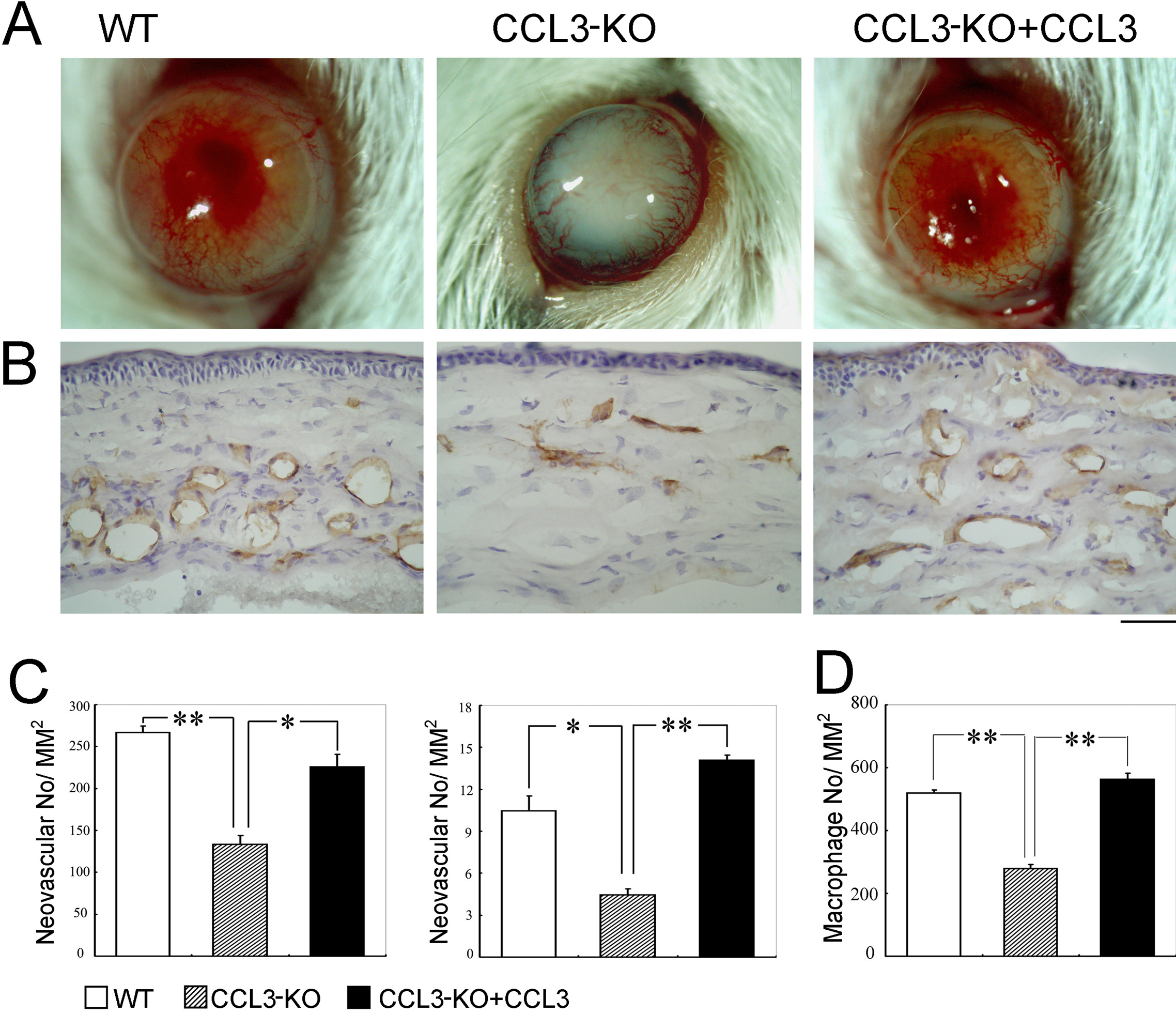
Figure 5. The effects of topical CCL3
application on corneal neovascularization. A: Macroscopic
appearances of WT, CCL3-KO mice, and CCL3-KO mice topically applied
with CCL3 two weeks after
alkali injury are shown.
Representative results from five animals from each group are shown
here. B: Corneal tissues were obtained two weeks after the
injury from WT, CCL3-KO, and CCL3-KO mice topically applied with CCL3
and were immunostained with anti-CD31 antibodies. Representative
results from five individual mice from each group are shown. Original
magnification, 400X. Scale bar, 50 μm. C: The CNV numbers per mm2
in hot spots (left panel) and % CNV areas in hot spots (right panel)
were determined. Each value represents the mean and SEM (n=5 animals). D:
The number of infiltrated F4/80 positive macrophages was determined on
WT, CCL3-KO, and CCL3-KO, which were all treated with CCL3, two days
after the injury. Each value mean represents both the mean and SEM
(n=5). The asterisk denotes a p<0.05, and the double asterisk means
a p<0.01 when compared with CCL3-KO (this applies to both C
and D).