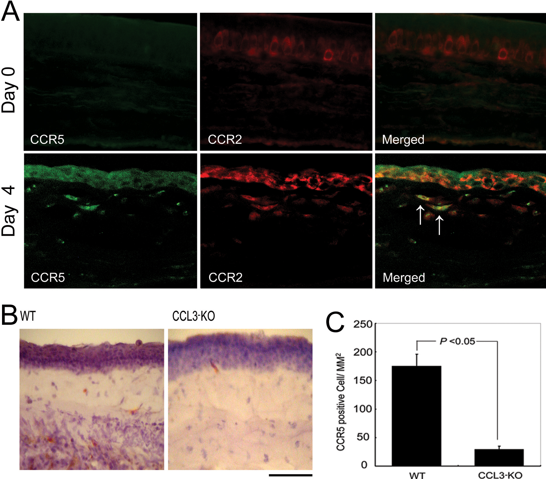
Figure 4. Intracorneal CCR5 positive cell
infiltration. A: A double-color immunofluorescence analysis of
CCR5-expressing cells is illustrated. Corneas were obtained from WT
mice 0 and 4 days after the injury. The samples were immunostained with
a combination of anti-CCR5 and anti-CCR2 antibodies as described in
Methods and observed with fluorescence microscopy (original
magnification, 400X). Signals were digitally merged in the right
panels. Arrows indicate the double, positively stained cells.
Representative results from three independent experiments are shown. B:
Corneal tissues from WT mice (left panel) or CCL3-KO mice (right panel)
obtained four days after the injury were stained with anti-CCR5 Ab.
Scale bar, 100 μm. C: The numbers of intracorneal CCR5 positive
cells four days after the injury were determined as described in
Methods, and the mean and SEM are shown here (n=5).