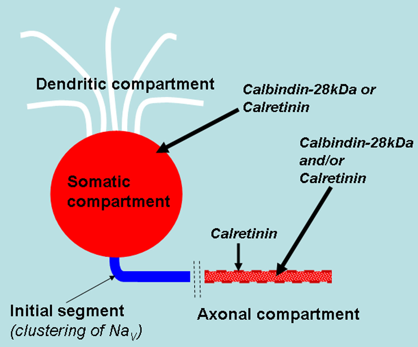
Figure 9. Schematic summarizing the
subcellular distribution of calbindin-28 kDa and calretinin in the
different compartments of the retinal ganglion cell. Labeling for
calbindin-28 kDa or calretinin was absent from the dendritic
compartment of the retinal ganglion cells (white). Many, but not all,
retinal ganglion cell soma show labeling for either calbindin-28 kDa or
calretinin (red). The initial segments of the retinal ganglion cells
where the voltage-gated sodium channels (NaVs in blue) are
clustered are not labeled by either calbindin-28 kDa or calretinin.
Calretinin immunolabeling is concentrated at distinct locations on the
retinal ganglion cell axon (dashed red line) where they are most likely
membrane bound. Calbindin-28 kDa and calretinin are either diffusely
coexpressed or differentially expressed in the axons (diffuse red
dots), but are both absent from the initial segments.