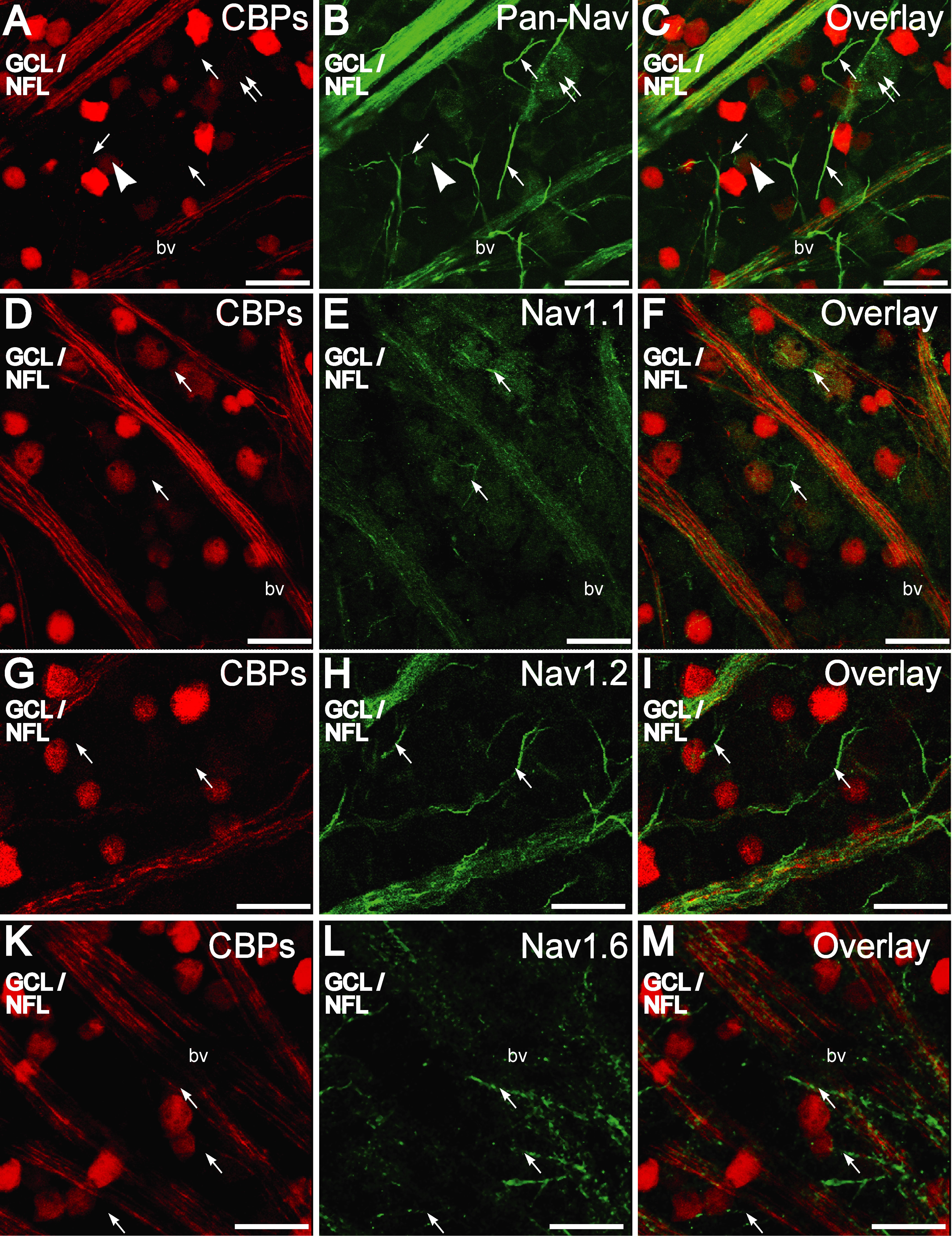
Figure 6. Comparison of immunofluorescence
for the calcium binding proteins, calretinin and calbindin-28 kDa, and
voltage-gated sodium channel antibodies in the ganglion cell layer
(GCL)/nerve fiber layer (NFL) as seen in retinal whole-mounts. A-C:
Calcium binding proteins (CBP) in the NFL are extensively colocalized
with Pan-NaV. Some Pan-NaV stained retinal ganglion cell
(RGC) somata were also immunopositive for CBPs (arrowhead) while others
were not (double arrows). Initial segments of RGCs, (arrow) some of
which can be seen emerging from the RGC somata, were immunopositive for
Pan-NaV but not colabeled with CBPs. D-F: NaV1.1-immunopositive
(green) RGC nerve fiber bundles in the nerve fiber layer (NFL) were
colabeled with CBPs (red), but the axon initial segments (arrow) were
not. G-I: NaV1.2 immunopositive (green) RGC nerve
fiber bundles in the NFL were colabeled with CBPs (red) but not the
axon initial segments (arrow). J-K: NaV1.6
immunopositive (green) axon initial segments (arrow) were not colabeled
with CBPs (red). Scale bar equals 20 µm. Abbreviations: bv is blood
vessel.