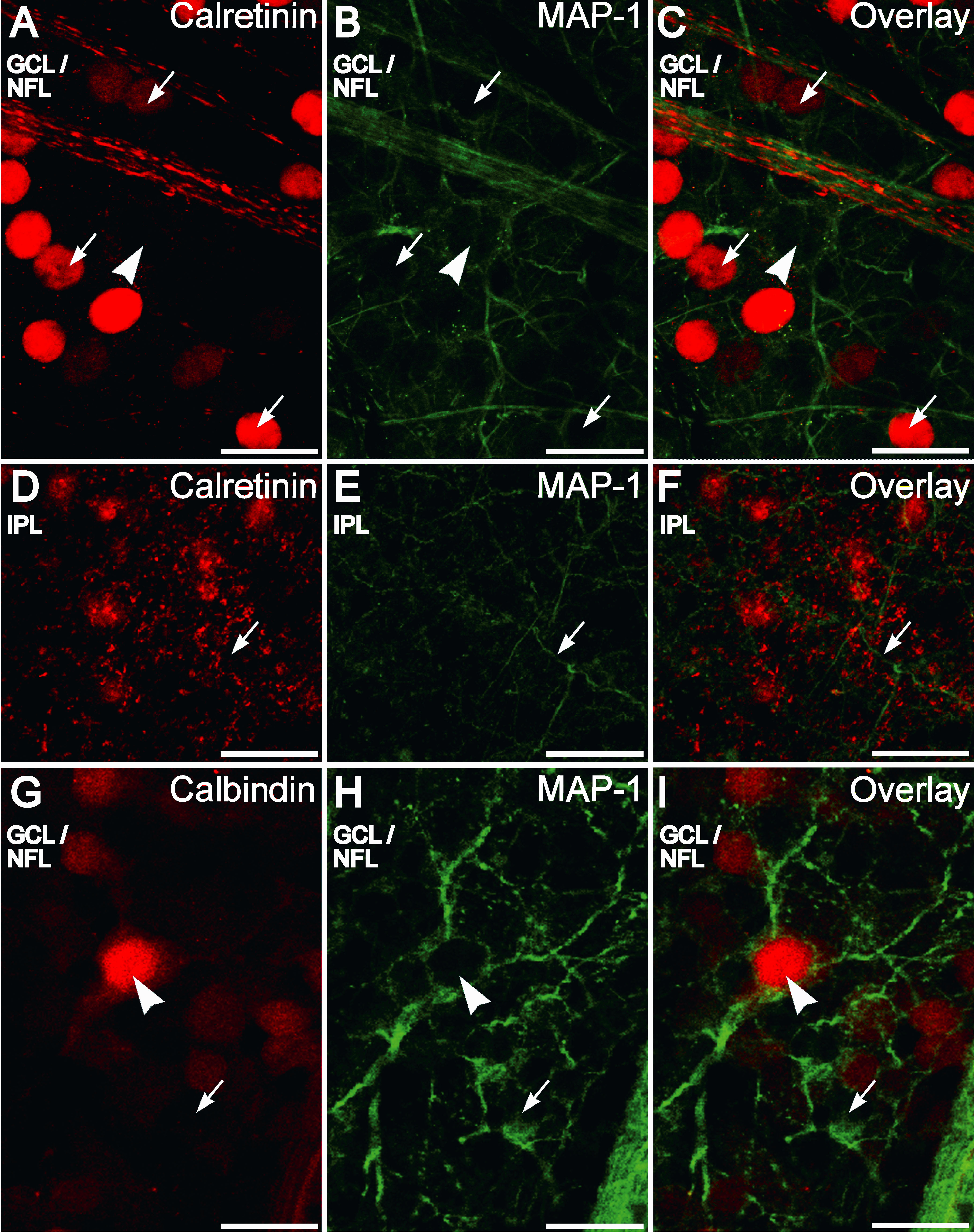
Figure 5. Calretinin and calbindin-28 kDa
immunofluorescence is not present in the distal dendritic compartment
of retinal ganglion cells as seen in retinal whole-mounts. A-C:
Double labeling for calretinin (red) and microtubule-associated protein
1 (MAP-1; green) in the ganglion cell layer (GCL)/nerve fiber layer
(NFL) shows that MAP-1 positive dendrites are not colabeled with
calretinin. Some large retinal ganglion cells (RGCs) that are
completely ringed by MAP-1 staining (arrowhead) are not positive for
calretinin. RGCs with smaller somata partially ringed with MAP-1
staining are immunopositive for calretinin (arrows). Other brightly
stained somata not showing MAP-1 immunofluorescence are the displaced
amacrine cells that also stained for calretinin. D-F: Confocal
plane showing that the MAP-1 positive (green) dendrites (arrow) do not
merge with the calretinin-positive plexus (red) in the inner plexiform
layer. G-I: Double labeling for calbindin-28 kDa (red) and
MAP-1 (green) in the GCL/NFL shows that some large RGCs that are
completely ringed by MAP1 staining (arrowhead) are positive for
calbindin-28 kDa. Some RGCs with smaller somata that are incompletely
ringed with MAP-1 are also calbindin-28 kDa positive while others are
not (arrow). Some neurons incompletely ringed with MAP-1 are also not
calbindin-28 kDa positive (arrow). Scale bar represents 20 µm.