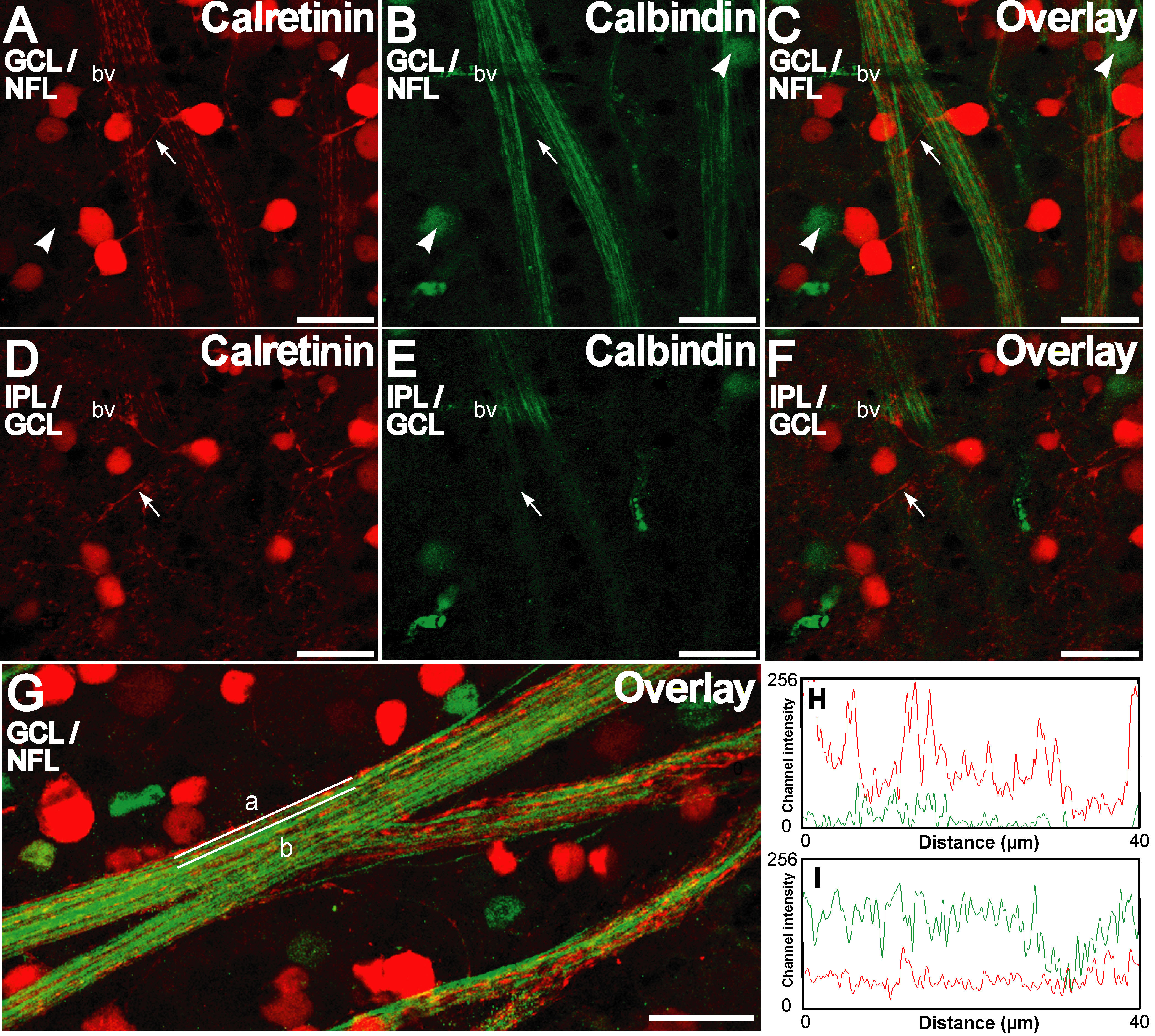
Figure 4. Calretinin and calbindin-28 kDa
are distinctly distributed in the ganglion cell and nerve fiber layer
as seen in retinal whole-mounts. A-C: Double labeling for
calretinin (red) and calbindin-28 kDa (green) in the ganglion cell
layer (GCL)/nerve fiber layer (NFL) shows that labeling for each was
present in a distinct set of neurons. Calbindin-28 kDa positive cell
bodies are indicated by arrowheads. Some calretinin-positive neurons
show processes (arrow) that ascend distally. Note the discontinuous
staining pattern of calretinin in the NFL in contrast to a smoother
staining pattern for calbindin-28 kDa. D,E: A single
confocal optical section distal to that of A-C shows that the
calretinin positive processes (arrow) are directed in the inner
plexiform layer (IPL) distally toward a calretinin-immunopositive
plexus, a characteristic of displaced amacrine cells. G:
Representative calretinin and calbindin-28 kDa double staining in the
GCL/NFL is shown. Channel intensity profiles for the red and green
channels for straight lines along the long axis (lines a and b in G
shown in H and I respectively) show different intensity
profiles for calretinin and calbindin-28kDa immunofluorescence. Scale
bar represents 20 µm.