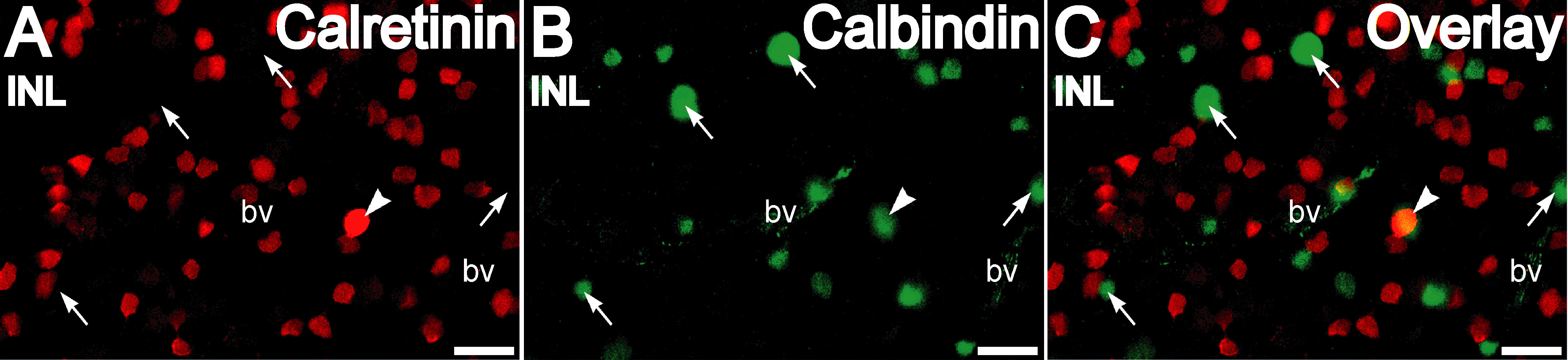
Figure 3. Calretinin and calbindin-28 kDa
are differentially distributed in amacrine cells as seen in retinal
whole-mounts. A-C: Double labeling for calretinin (red) and
calbindin-28 kDa (green) in the inner nuclear layer shows that labeling
for each was present in a distinct set of amacrine cells For the
apparent region of overlap (arrowhead) in the overlay of this confocal
plane, examination of different z-planes revealed that these were
disparate cells located at different depths. Arrows indicate
calbindin-28 kDa-immunopositive cells. Scale bar represents 20 µm.
Abbreviations: bv is blood vessel.