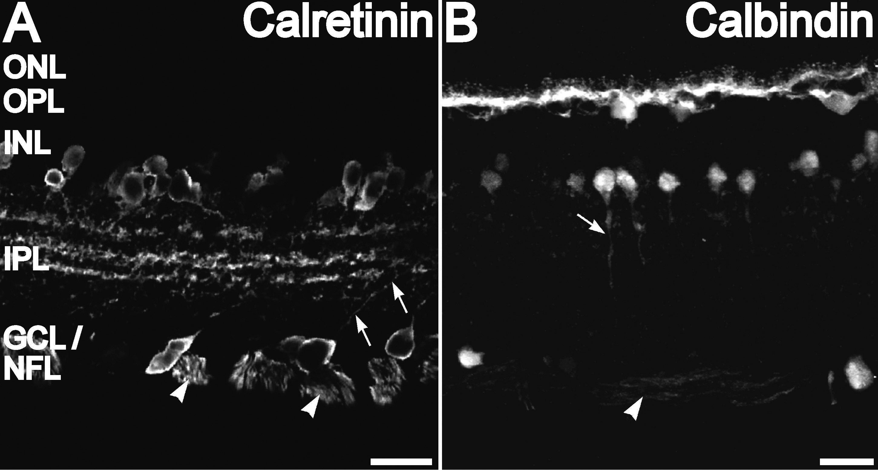
Figure 2. Calretinin and calbindin-28 kDa
immunolabeling of a vertical cryosection. A: Calretinin
immunolabeling was present in cell bodies and processes of amacrine
cells at the inner nuclear layer (INL)-inner plexiform layer (IPL)
border. Calretinin labeling was also present in cell bodies and
processes (arrows) in the ganglion cell layer (GCL). Calretinin
labeling is also found in three distinct bands in the IPL and retinal
ganglion cell (RGC) axons in the nerve fiber layer (NFL; arrowhead). B:
Calbindin-28 kDa immunolabeling was present in cell bodies and
processes of horizontal cells at the outer plexiform layer (OPL)-INL
border. Calbindin-28 kDa also labeled amacrine cells at the INL-IPL
border. Some descending processes were seen for some of these neurons
(arrow). There are also diffuse calbindin-28 kDa-positive punctate in
the IPL. Calbindin-28 kDa labeling is seen in few cells in the GCL as
well as in the RGC axons in the NFL (arrowhead). Scale bars represent
20 µm.