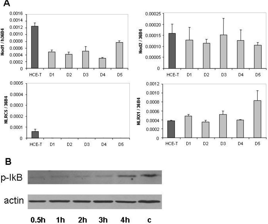
Figure 3. Expression of Nod subtypes in
corneal epithelial cells.
A: The relative expression of
Nod1,
Nod2,
NLRC5, and
NLRX1 mRNA in HCE-T cells and
in primary corneal epithelial cells derived from five individuals were
measured and documented as described in the legend of
Figure 1.
B:
Detection of phosphorylated IκB using western blotting is illustrated.
HCE-T cells were treated with 4 μg/ml ultrapure PGN-ECndss for the
indicated time periods. The control THP-1 cells (c) were treated with 1
μg/ml LPS for 4 h. Equal amount of sample loading was verified by
detecting β-actin protein expression.