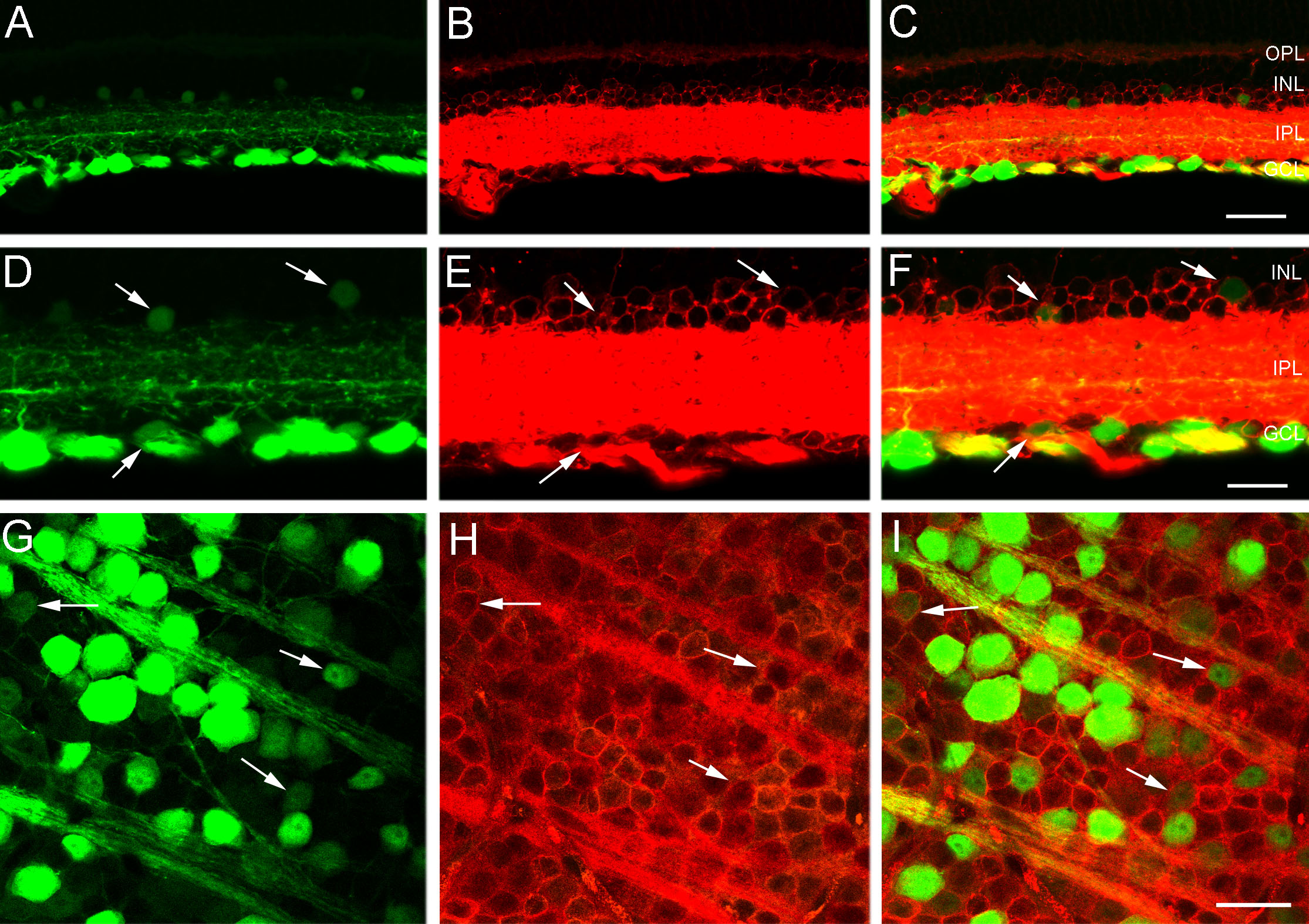
Figure 6. Small, weak CFP fluorescent
somata in the INL and GCL contain HPC-1 immunoreactivity, a marker of
amacrine and displaced amacrine cells. A: A transverse section
of peripheral retina shows cyan fluorescent protein (CFP) expression in
numerous somata in the ganglion cell layer (GCL) and weak
CFP-expressing somata in the proximal inner nuclear layer (INL). B:
Same section as in A shows syntaxin 1a (HPC-1) immunoreactivity
in amacrine and displaced amacrine cell somata in the INL and GCL,
respectively. HPC-1 immunostaining is very strong in the inner
plexiform layer (IPL) and the weaker HPC-1 immunoreactive somata in the
GCL tend to be obscured in transverse sections. C: A merged
image of A and B shows colocalization of CFP and HPC-1
immunoreactivity in small, weakly CFP-expressing cell bodies in the INL
and GCL. The scale bar for A-C is 40 μm. D: A
higher magnification image of transverse retina shows CFP fluorescence
in ganglion cells in the GCL and in smaller, weakly CFP fluorescent
cells in the INL and GCL (arrows). E: The same section as in D
shows HPC-1 immunoreactivity in numerous amacrine and displaced
amacrine cell somata in the INL and GCL (arrows). F: A merged
image of D and E shows colocalization of CFP expression
and HPC-1 immunoreactivity in amacrine and displaced amacrine cells
(arrows) in the INL, and GCL. The scale bar for D–F is 25 μm. G:
CFP is localized to brightly and weakly fluorescent cell bodies of
various sizes in the GCL. Image is from a retinal wholemount located
1.5 mm from the optic nerve head in midperipheral nasal retina. H:
Same region as in G shows HPC-1 immunoreactivity in numerous
cell somata in the GCL. I: A merged image of G and H
shows the colocalization of CFP and HPC-1 immunoreactivity in displaced
amacrine cell bodies in the GCL (arrows). The scale bar for G-I
is 30 μm. In C and F outer plexiform layer is
abbreviated outer plexiform layer (OPL).