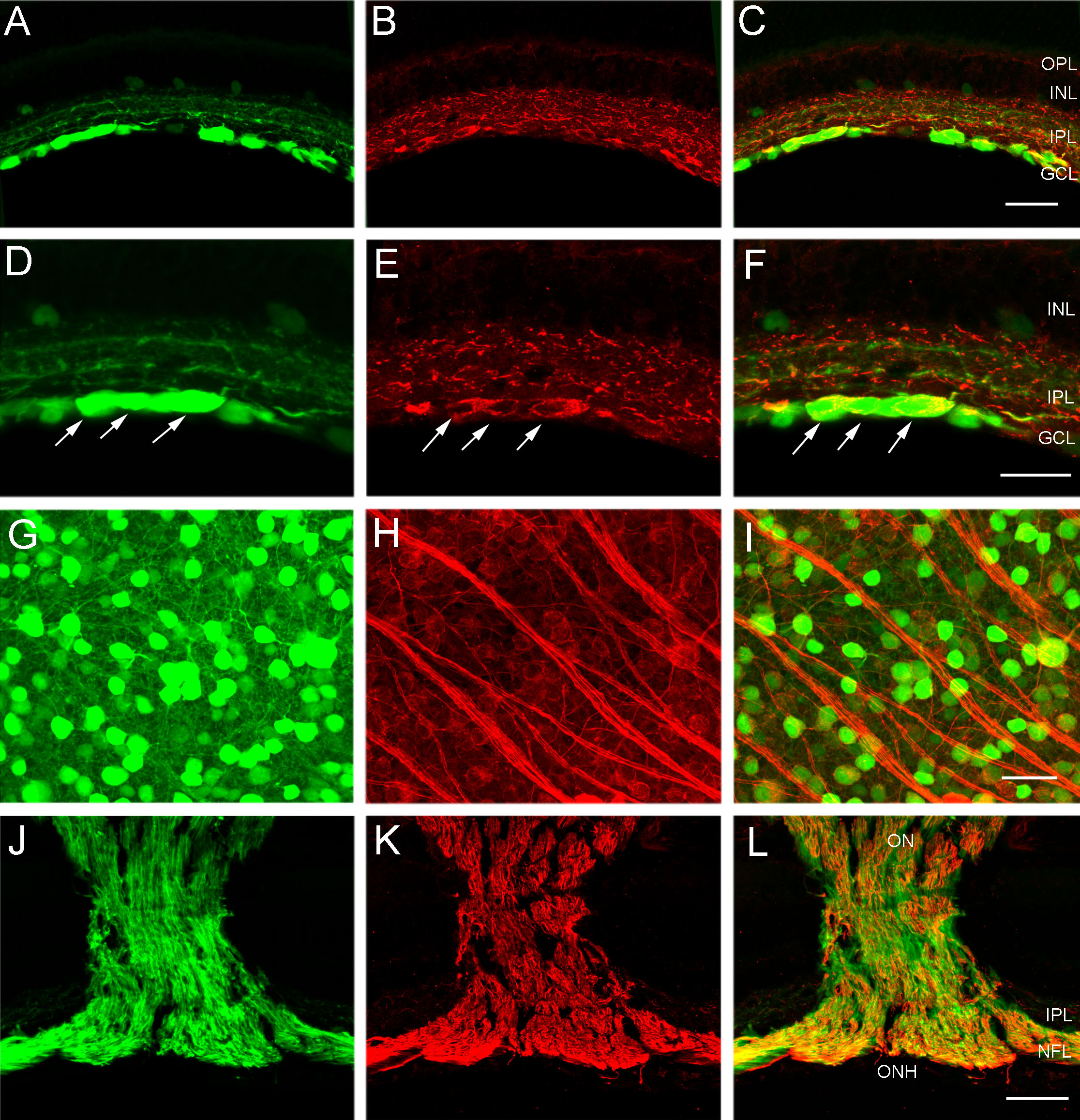
Figure 4. CFP-containing ganglion cells
express NF-L immunoreactivity in their soma, dendrites and axons in the
fiber layer, optic nerve head and optic nerve. A: Transverse
section of peripheral retina shows cyan fluorescent protein (CFP)
expression in numerous cell bodies in the ganglion cell layer (GCL). B:
Same section as in A shows neurofilament light (NF-L)
immunoreactivity in ganglion cell somata in the GCL and dendrites in
the inner plexiform layer (IPL). C: A merged image of A
and B demonstrates colocalization of CFP expression and NF-L
immunoreactivity in many cell bodies in the GCL and dendrites in the
IPL. The scale bar for A-C is 40 μm. D: A higher
magnification image of transverse retina shows CFP fluorescence in
ganglion cells, including large ganglion cell somata (arrows). E:
NF-L immunoreactivity is evident in numerous cell bodies in the GCL
including the large ganglion cell somata (arrows). F: A merged
image of D and E demonstrates colocalization of CFP
expression and NF-L immunoreactivity in most cell bodies in the GCL and
dendrites in the IPL. The scale bar for D-F is 25 μm. G:
CFP is localized to brightly and weakly fluorescent cell bodies of
various sizes in the GCL. Image is from a retinal wholemount located
1.5 mm from the optic nerve head in midperipheral nasal retina. H:
NF-L immunoreactivity is evident in ganglion cell somata in the same
region as G. I: A merged image of G and H
shows colocalization of CFP expression and NF-L immunoreactivity in
ganglion cell bodies. The scale bar for G-I is 30 μm. J:
CFP fluorescence is evident in ganglion cell axons in the fiber layer,
optic nerve head and optic nerve, in this low magnification image of a
transverse section through the optic nerve head. K: The same
section as in J, shows NF-L immunoreactivity in ganglion cell
axons. L: A merged image of J and K shows
colocalization of CFP and NF-L immunoreactivity in most ganglion cell
axons in the fiber layer, optic nerve head and optic nerve. Note: there
is not complete overlap of CFP and NF-L in ganglion cell axons, as some
NF-L labeled cells do not express CFP. The scale bar for J–L is
140 μm. In C and F inner nuclear layer is abbreviated
INL, and outer plexiform layer is abbreviated OPL.