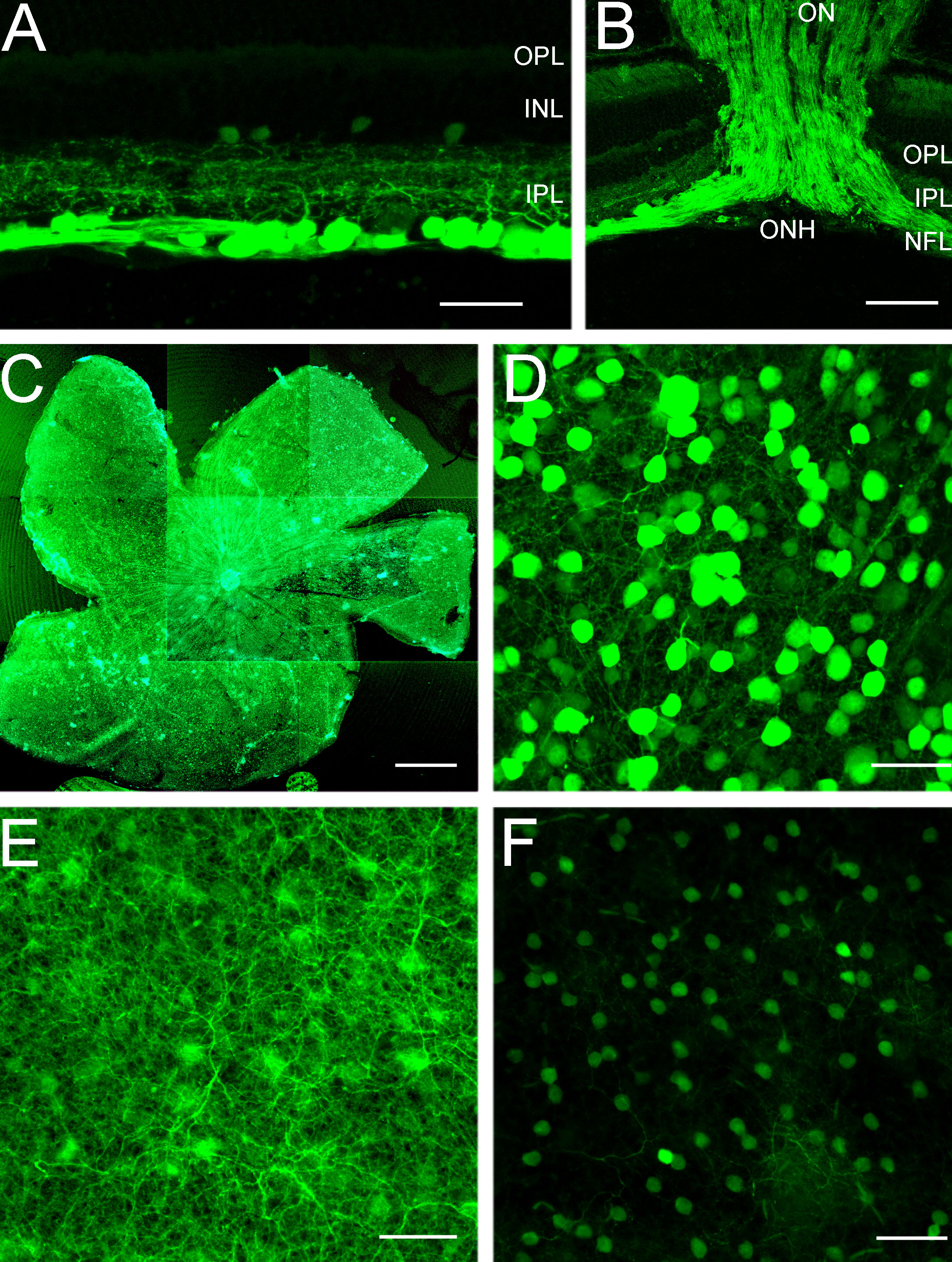
Figure 1. Cyan fluorescent protein
expression in the retinas of thy1-CFP transgenic mice in
confocal images of transverse sections (A,B) and wholemounts (C-F).
A: Most cyan fluorescent protein (CFP) expression is localized
to brightly fluorescent cell bodies in the ganglion cell layer (GCL),
processes that form a dense plexus in all laminae of the inner
plexiform layer (IPL) and axons in the nerve fiber layer (NFL). Small,
weakly CFP fluorescent cells are in the proximal inner nuclear layer
(INL) and the GCL. The scale bar represents 35 µm. B: CFP
expression is prominent in ganglion cell axons in the NFL, optic nerve
head (ONH), and optic nerve (ON). Scale bar equals 60 µm. C:
Low-magnification composite image of a wholemount shows CFP expression
in all retinal regions. Scale bar equals 550 μm. D: CFP
expression is localized to brightly and weakly fluorescent cell bodies
of various sizes in the GCL. Image is taken in midperipheral nasal
retina, 1.5 mm from the optic nerve head. E: A plexus of
CFP-containing processes occupies the IPL. Figure E shows the same
region as in D. F: Weak CFP expression is evident in
small cell bodies in the proximal INL. Panel F shows the same
region as in D. Scale bar for D-F equals 35 μm.
In A and B, outer plexiform layer is abbreviated OPL.