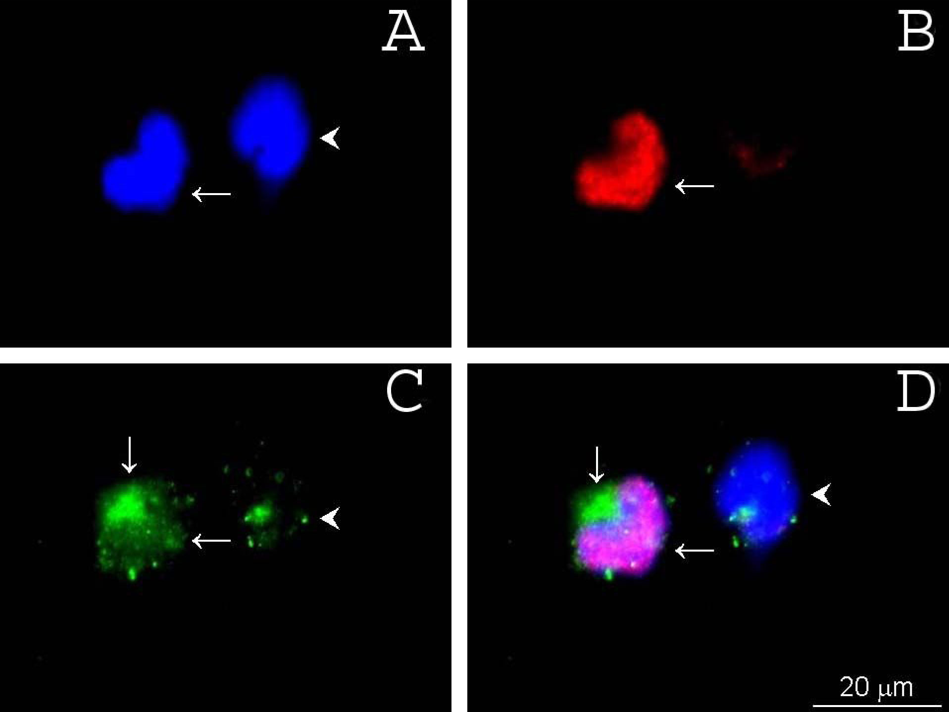
Figure 7. γ-Synuclein subcellular
localization was examined using immunofluorescent staining of rat
retinal cells. A: DAPI (blue) counterstained the nuclei. B:
Brn-3a (red) was localized to RGC nuclei. C: γ-synuclein
(green) was co-expressed by the Brn-3a expressing cell (arrows) but
only weakly by a non-Brn-3a-expressing cell (arrowhead). A merged image
of A, B, and C is shown in D.
Immunofluorescent staining of retinal cell suspension. A: DAPI,
B: Brn-3a, C: γ-synuclein, D: merged images.
Horizontal arrows show a cell immunopositive for both Brn-3a and
γ-synuclein. Arrowhead (C and D) points to a cell not
stained by Brn-3a antibody that is stained by a γ-synuclein antibody.
Vertical arrow (C and D)points to the
localization of γ-synuclein in the perinuclear area. Thus, brighter and
broader cytoplasmic staining of γ-synuclein overlapped well with Brn-3a
staining.