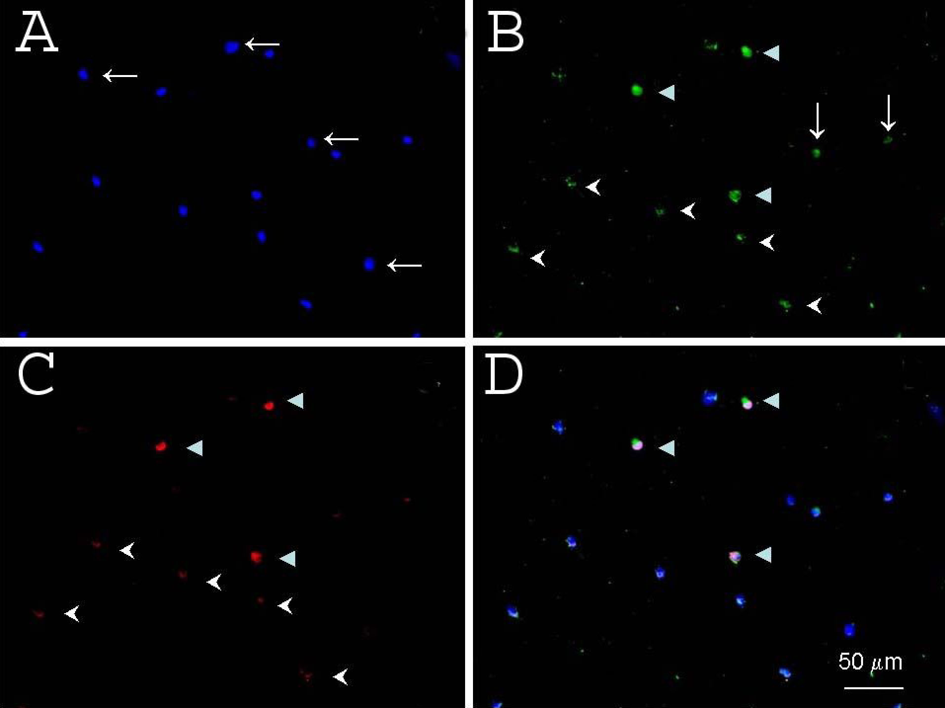
Figure 6. γ-Synuclein specificity was
examined using immunofluorescent staining of rat retinal cells. A:
DAPI (blue) counterstained the nuclei. B: Brn-3a (green) was
localized to RGC nuclei. C: γ-synuclein (red) was localized
throughout the cytoplasm (arrowheads) and processes (arrows). A merged
image of A, B, and C is shown in D.
Many cells demonstrated neither Brn-3a nor γ-synuclein reactivity
(horizontal arrows in A). A subset of presumptive retinal
ganglion cells demonstrated intensive staining by both γ-synuclein and
Brn-3a (full arrowheads in B, C, and D), while
many other cells showed only weak staining by both γ-synuclein and
Brn-3a (arrowheads in B and C). A few cells were
stained by γ-synuclein, but not by Brn-3a (vertical arrows in B).