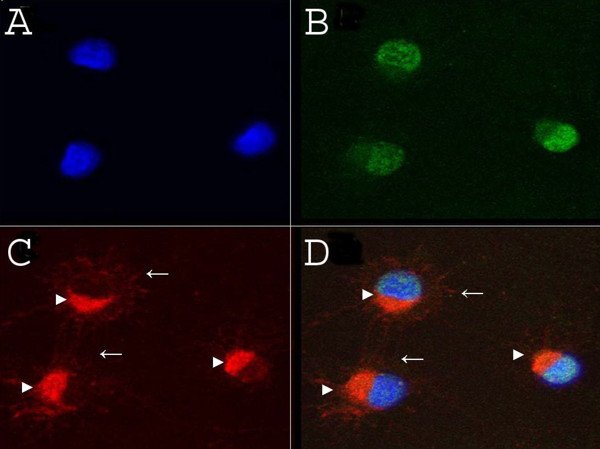
Figure 5. Localization of γ-synuclein and
Brn-3a were examined in a primary culture of rat retinal ganglion cells
(RGCs). A: DAPI (blue) counterstained the nuclei. B:
Brn-3a (green) was localized to RGC nuclei. C: γ-synuclein
(red) was localized throughout the cytoplasm (arrowheads) and
processes. A merged image of A, B, and C is shown in D.
Thus, in primary culture of RGCs, γ-synuclein is localized in the
cytoplasm and processes, while Brn-3a in the nuclei.