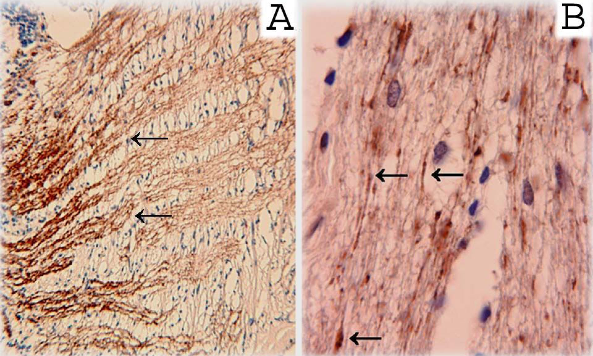
Figure 3. Immunohistochemical staining
with a γ-synuclein antibody was used to examine a non-glaucomatous
human optic nerve in the area immediately posterior to the lamina
cribrosa (A) and the retrobulbar optic nerve from a patient with
primary open-angle glaucoma (B). In both sections, γ-synuclein
reactivity can be seen along presumptive retinal ganglion cell axon
bundles. In section B, arrows show swollen axons and axons
fragment immunopositive for γ-synuclein. These results confirm the
presence of γ-synuclein in axons of RGC and its immunopathology in
glaucoma.