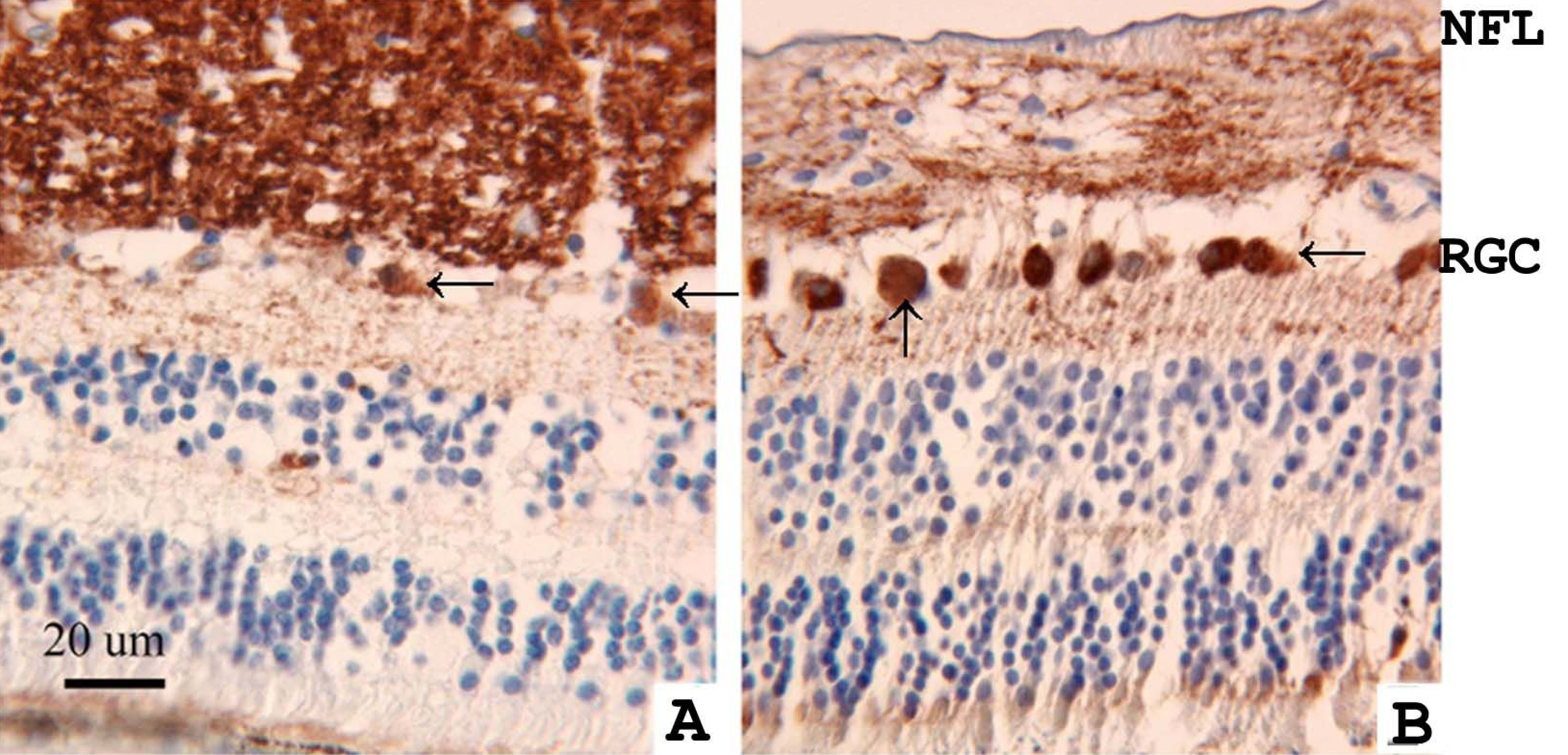
Figure 2. Localization of γ-synuclein was
studied by immunohistochemistry in a normal human retina (A) and
in the retina of an age-matched patient with retinoblastoma (B).
γ-Synuclein was detected using a diaminobenzidine reagent (brown,
peroxidase). Tissues were counterstained with hematoxylin (blue).
Sections were stained with an antibody that recognized γ-synuclein, but
not the other members of the synuclein family. The retina of the
retinoblastoma patient (B) demonstrated more intense γ-synuclein
immunoreactivity in retinal ganglion cell bodies (arrows) and less
intensive staining in the nerve fiber layer (NFL) compared with the
normal retina (A).