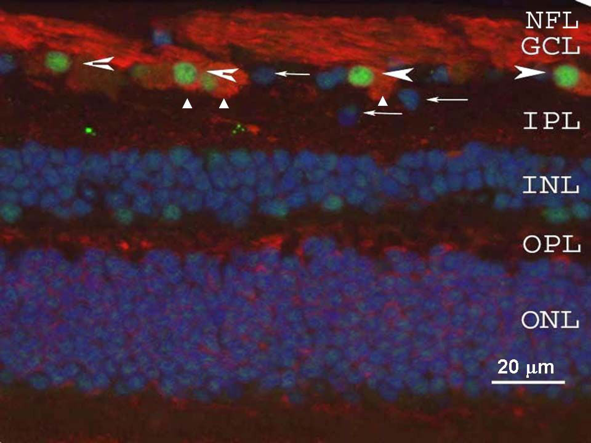
Figure 1. γ-Synuclein and Brn-3a
localization were examined in the human retina by immunofluorescence.
γ-Synuclein (red) was present in the cytoplasm of retinal ganglion
cells (RGCs; triangles) and in the nerve fiber layer (NFL), while
Brn-3a (green, arrowheads) was observed in RGC nuclei. Arrows mark
cells in the retinal ganglion cell layer (GCL) that were not stained by
either Brn-3a or γ-synuclein. Nuclei throughout the retina are
counterstained with DAPI (blue), and little immunofluorescence is noted
through the inner or outer plexiform layers (IPL and OPL,
respectively), or inner and outer nuclear layers (INL and ONL,
respectively). Thus γ-synuclein is localized both in the body of RGC
and in their axons.