Figure 4 of
Zhuo, Mol Vis 2008; 14:1533-1539.
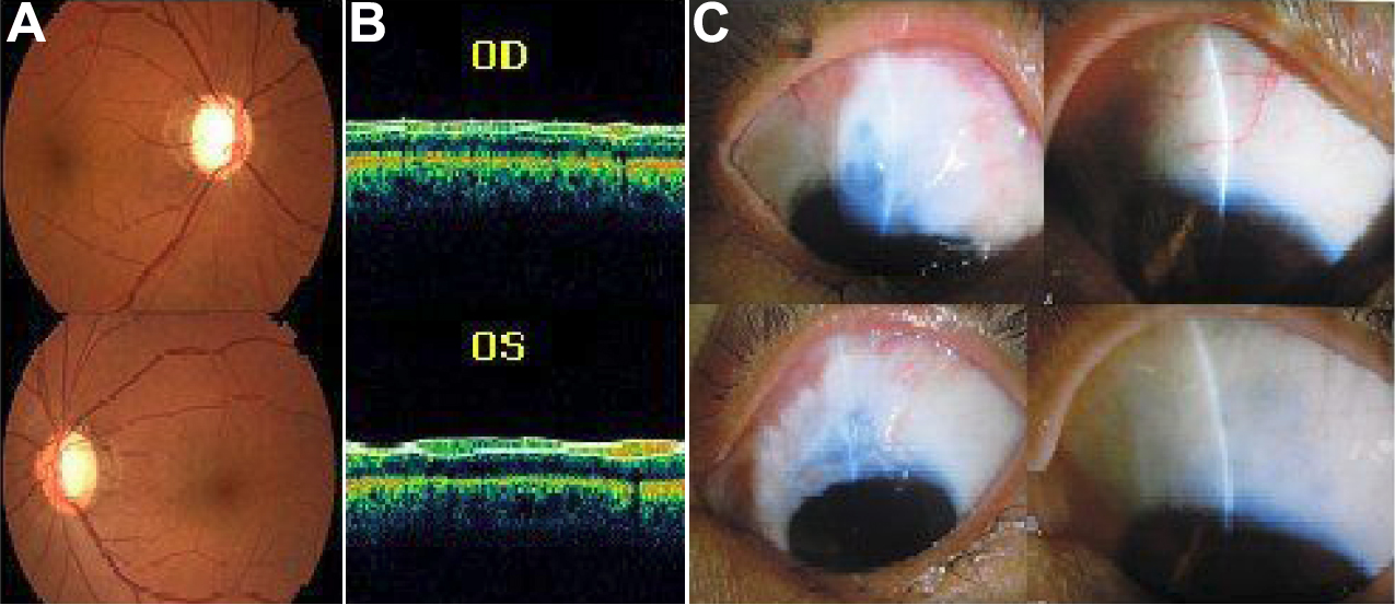
Figure 4.
Individual III5 (top) right eye, (bottom) left eye.
A
: Fundus images showing typical glaucomatous cupping of the optic disc,
B
: OCT images showing thinner nerve fiber layer,
C
: Thin-walled filtering blebs after filtering surgery.