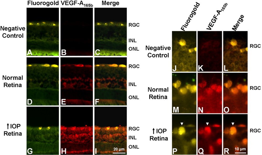
Figure 5. Immunohistochemical analysis of
VEGF-A165b expression in the glaucomatous retina after five
days of elevated IOP. A-C: Negative control.
Non-specific staining was not detected in the retina. D-F:
VEGF-A165b staining of the normal retina (n=4). VEGF-A165b
was present in the RGC and the INL. G-I: VEGF-A165b
staining of the glaucomatous retina (n=4). VEGF-A165b
staining was stronger in the RGC and the INL compared to the normal
retina. J-L: Negative control. No non-specific staining
was observed in the retina. M-O: VEGF-A165b
staining of the RGC in the normal retina. Staining was colocalized with
the RGC marker, Fluorogold. P-R: VEGF-A165b
staining of the RGC in the glaucomatous retina. Levels of VEGF-A165b
in the Fluorogold-labeled RGC (white arrow) were increased in the
retinas with elevated IOP.