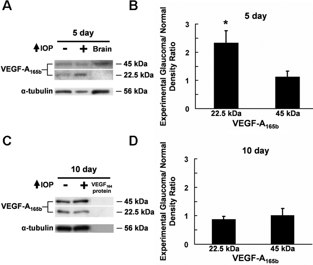
Figure 4. Western blot analysis of VEGF-A165b
expression in the glaucomatous retina. A: VEGF-A165b
expression following five days of elevated IOP. Retinal VEGF-A165b
monomer and dimer were detected at 22.5 and 45 kDa, respectively. B:
Glaucomatous/control ratio of normalized VEGF-A165b
densitometry readings in the retina following five days of elevated
IOP. Expression of the 22.5 kDa VEGF-A165b was increased
significantly in the glaucomatous retinas compared to the controls. C:
VEGF-A165b expression following 10 days of elevated IOP.
VEGF-A165b monomer and dimer were observed at 22.5 and 45
kDa in the retina, respectively. D: Glaucomatous/control ratio
of normalized VEGF-A165b densitometry readings in the retina
following 10 days of elevated IOP. Both 22.5 kDa and 45 kDa VEGF-A165b
were expressed at comparable levels in the control and glaucomatous
retinas. The positive control was the brain while the negative control
was VEGF-A164 recombinant protein. The loading control was
α-tubulin.