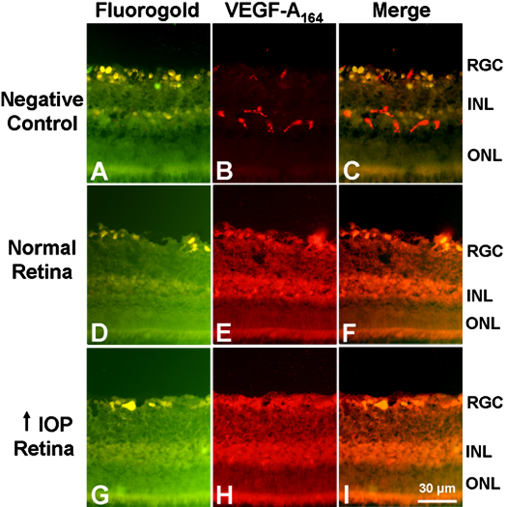
Figure 2. Immunohistochemical analysis of
VEGF-A164 expression in the glaucomatous retina after five
days of elevated IOP. A-C: Negative control. Some
non-specific staining of blood vessels in the RGC and the INL was
observed. D-F: VEGF-A164 staining of the
normal retina (n=4). VEGF-A164 was present in the RGC and
the INL. G-I: VEGF-A164 staining of the
glaucomatous retina (n=4). Staining was detected in the RGC and INL.
VEGF-A164 levels did not differ between the normal and
glaucomatous retinas.