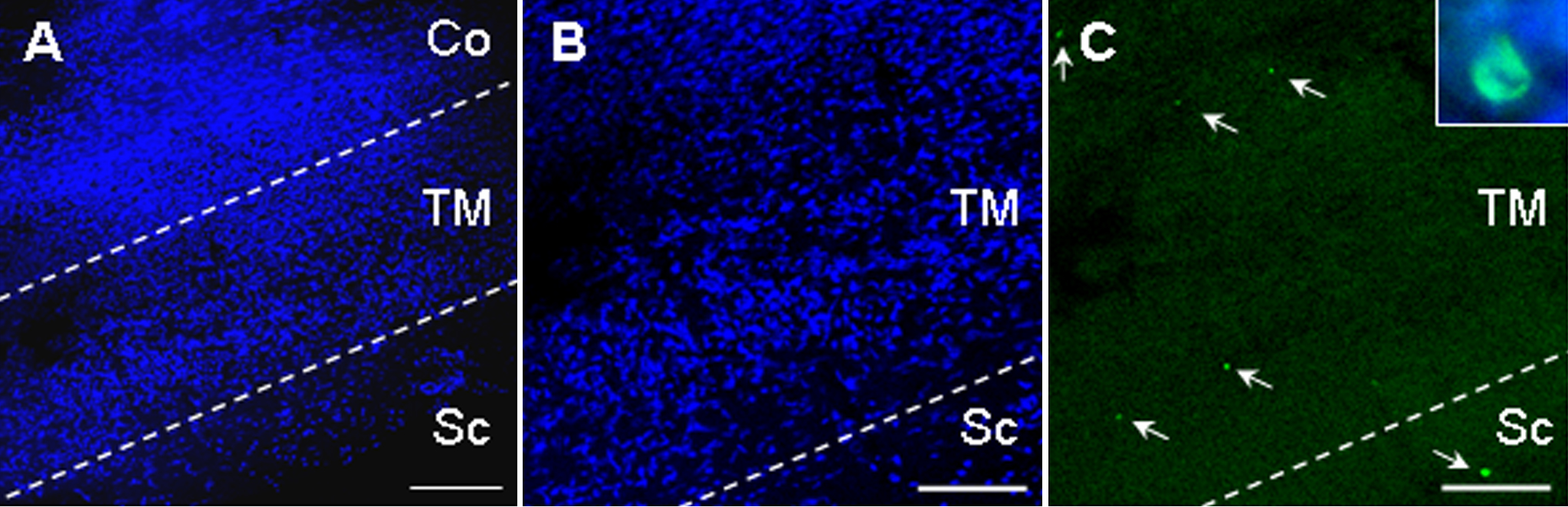
Figure 1. TUNEL positive cells can be
detected in TM samples from glaucomatous patients. TM samples were
stained with DAPI (A,B) and by TUNEL (C). The DAPI
staining allowed us to visualize the structure of the specimen and to
identify apoptotic nuclei both within and outside of the TM. Dotted
lines represent the limits of the TM and the arrows indicate the
presence of apoptotic cells in the TM and sclera of a POAG patient. The
inset in C shows a pyknotic nucleus (visualized by
DAPI blue staining) that is also positive for TUNEL (green).
Abbreviations in the figure are Co, Cornea; TM, Trabecular Meshwork;
Sc, Sclera. The scale bar is equal to 250 μm in A and 150 μm in
B and C.