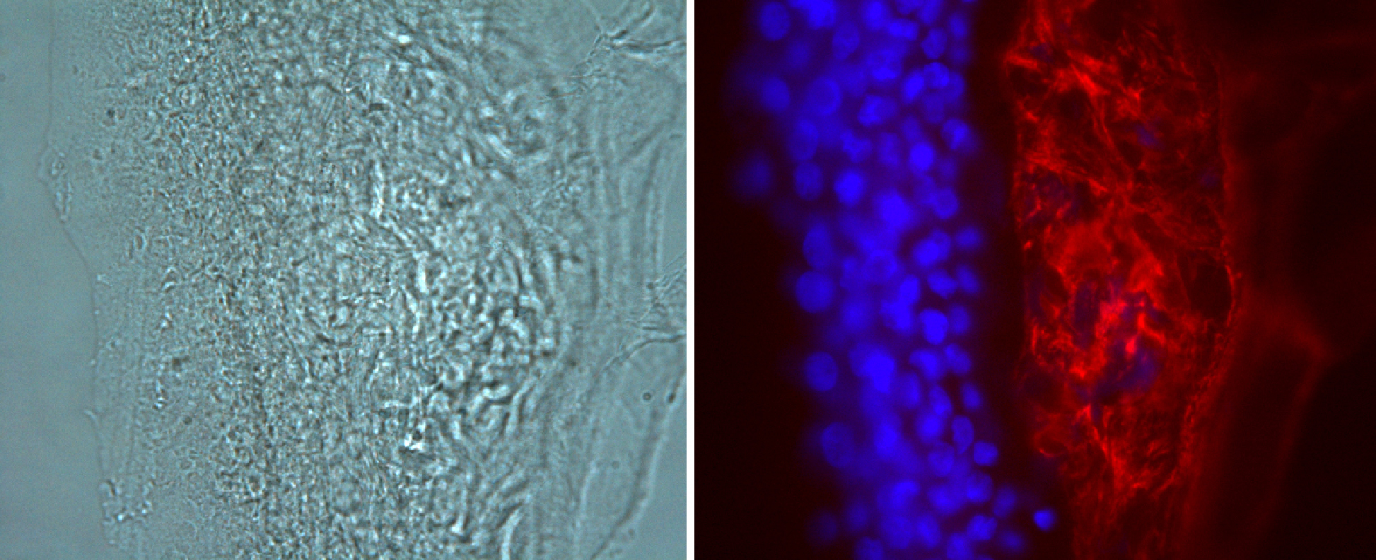
Figure 5. Immunohistochemical staining of
a corneal allograft specimen (II.1). The presence of TGFBI associated
protein is demonstrated in the sub epithelial deposit (red). Corneal
epithelial nuclei are stained with DAPI (blue). The right hand image
shows the corresponding phase contrast light microscopy image.
Magnification 40X.