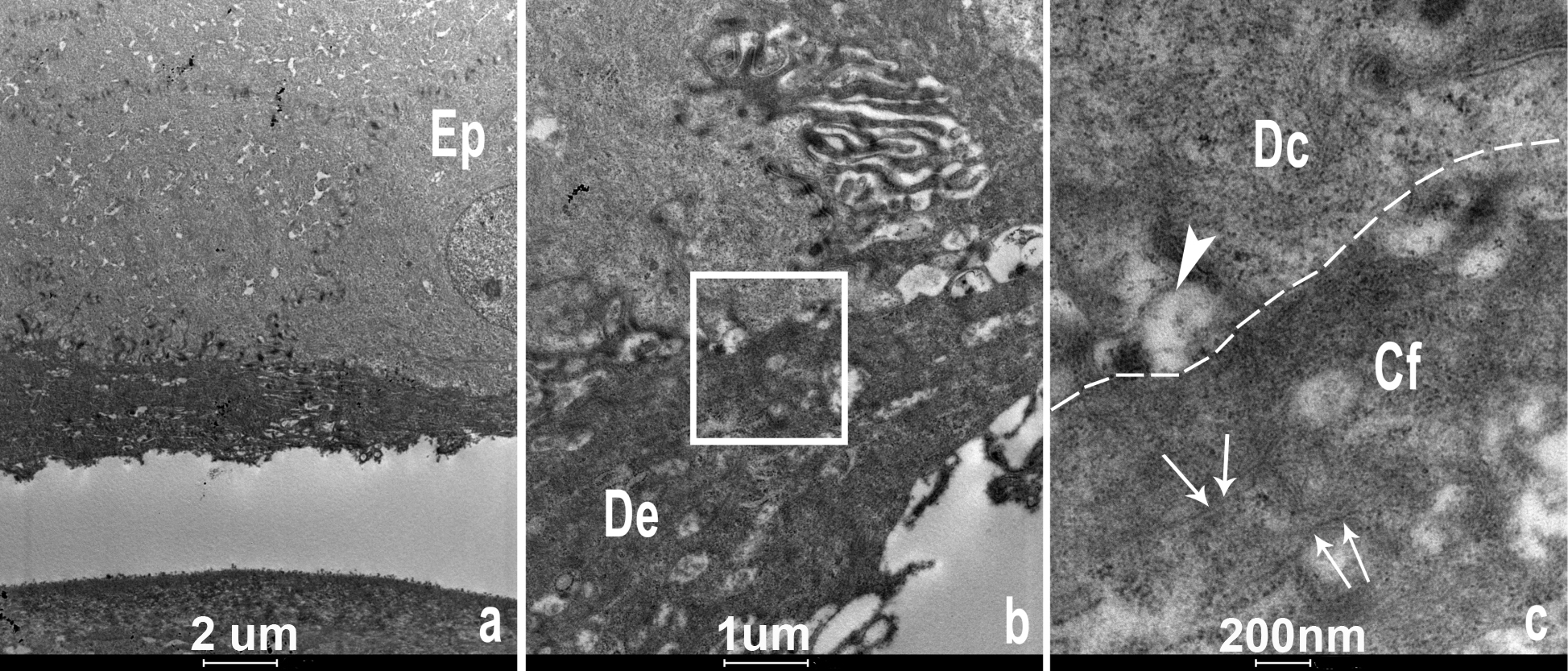
Figure 4. Transmission electron microscopy
images of the corneal allograft from subject II:1. A: Low
magnification photomicrograph showing basal epithelial cells (Ep)
overlying a band of deposit (De), at higher magnification in B.
The insert in B is shown in higher magnification in C,
demonstrating the degenerating basal epithelial cell (Dc) above the
dotted line, and below the subepithelial deposit, consisting of
irregular aggregates of fibrils (Cf) as demonstrated with arrows. The
large arrowhead shows a degenerating adhesion body.