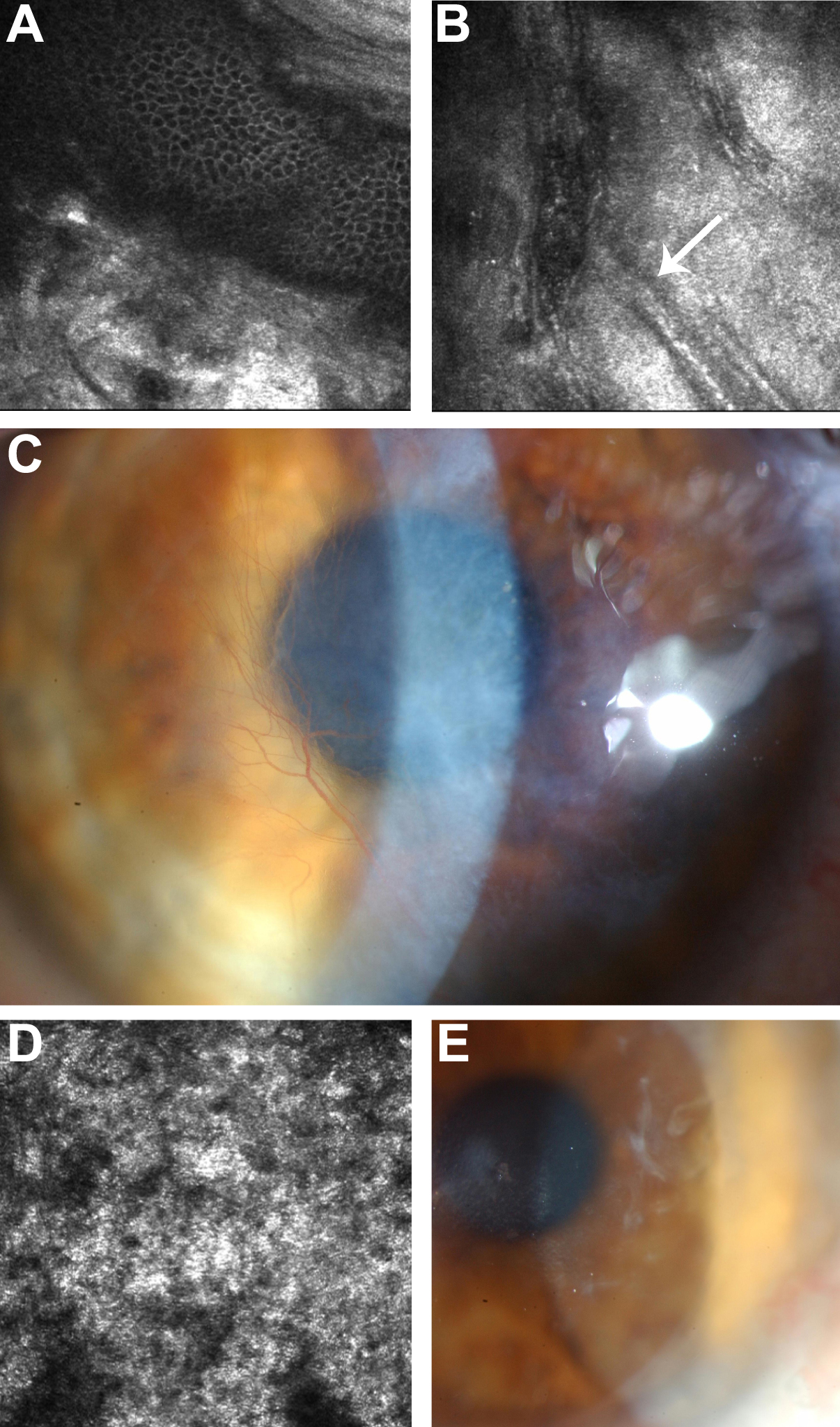
Figure 3. In vivo confocal microscopy
images of representative affected family members. The right eye (virgin
cornea) of the proband (frame size 400 µm x 400 µm) at the level of (A)
basal epithelial layer illustrating focal deposition of homogeneous,
reflective materials with rounded edges, and hyporeflective borders and
(B) posterior stroma showing extensive scarring with arrow
indicating “ghost” blood vessels. C: Corresponding slit-lamp
biomicroscopy photograph illustrating dense scarring. D: In
vivo confocal microscopy of the allograft in the right eye of subject
II:1 at the level of Bowman’s layer which has been completely replaced
by diffuse, homogeneous, reflective material (frame size 400 µm x 400
µm). E: Corresponding slit-lamp biomicroscopy photograph
illustrating recurrence of the dystrophy within the peripheral right
corneal graft of subject II:1, 4 years following penetrating
keratoplasty.

![]() Figure 3 of Wheeldon,
Mol Vis 2008; 14:1503-1512.
Figure 3 of Wheeldon,
Mol Vis 2008; 14:1503-1512. 