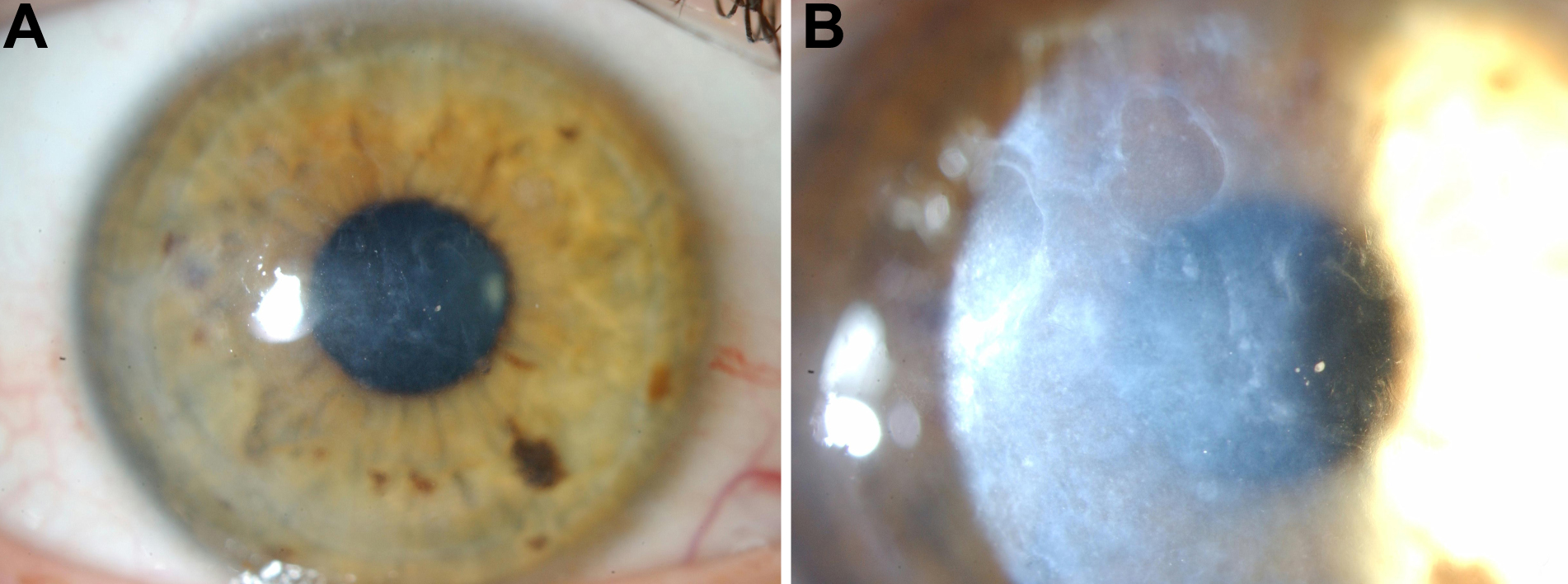
Figure 2. Representative slit-lamp
biomicroscopy photographs illustrating corneal phenotype in an affected
individual. A: 10X magnification diffuse illumination and B:
16X magnification broad slit lamp slit illumination of the left cornea
of subject III:5.

![]() Figure 2 of Wheeldon,
Mol Vis 2008; 14:1503-1512.
Figure 2 of Wheeldon,
Mol Vis 2008; 14:1503-1512. 