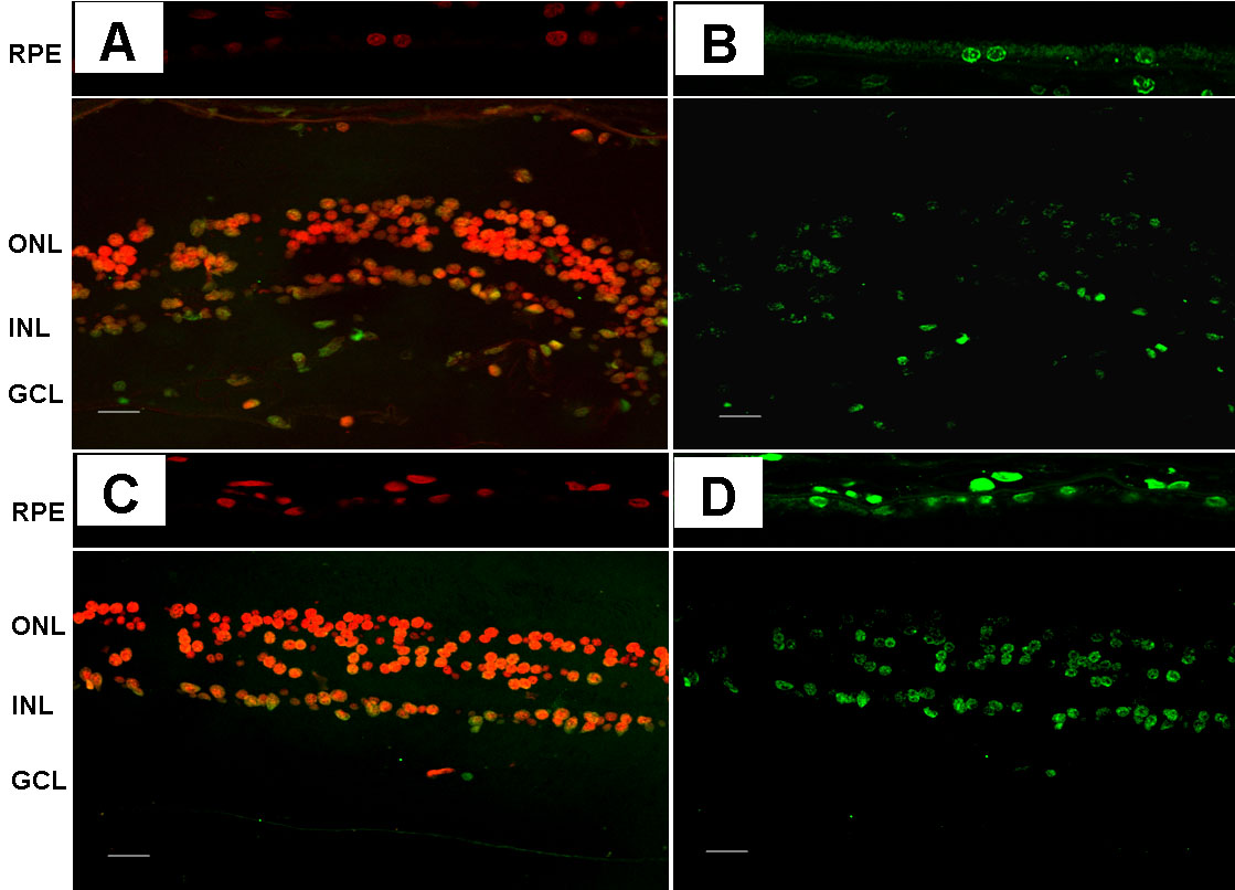
Figure 3. Representative images of
apoptosis in the retina. Upper panel: nondiabetic donor (A:
propidium iodide, B: TUNEL immunofluorescence). Lower panel:
diabetic donor (C: propidium iodide, D: TUNEL
immunofluorescence). The following abbreviations are used in the
figure: retinal pigment epithelium (RPE); outer nuclear layer (ONL);
inner nuclear layer (INL); and ganglion cell layer (GCL). The bar
represents 20 μm.

![]() Figure 3 of Carrasco,
Mol Vis 2008; 14:1496-1502.
Figure 3 of Carrasco,
Mol Vis 2008; 14:1496-1502. 