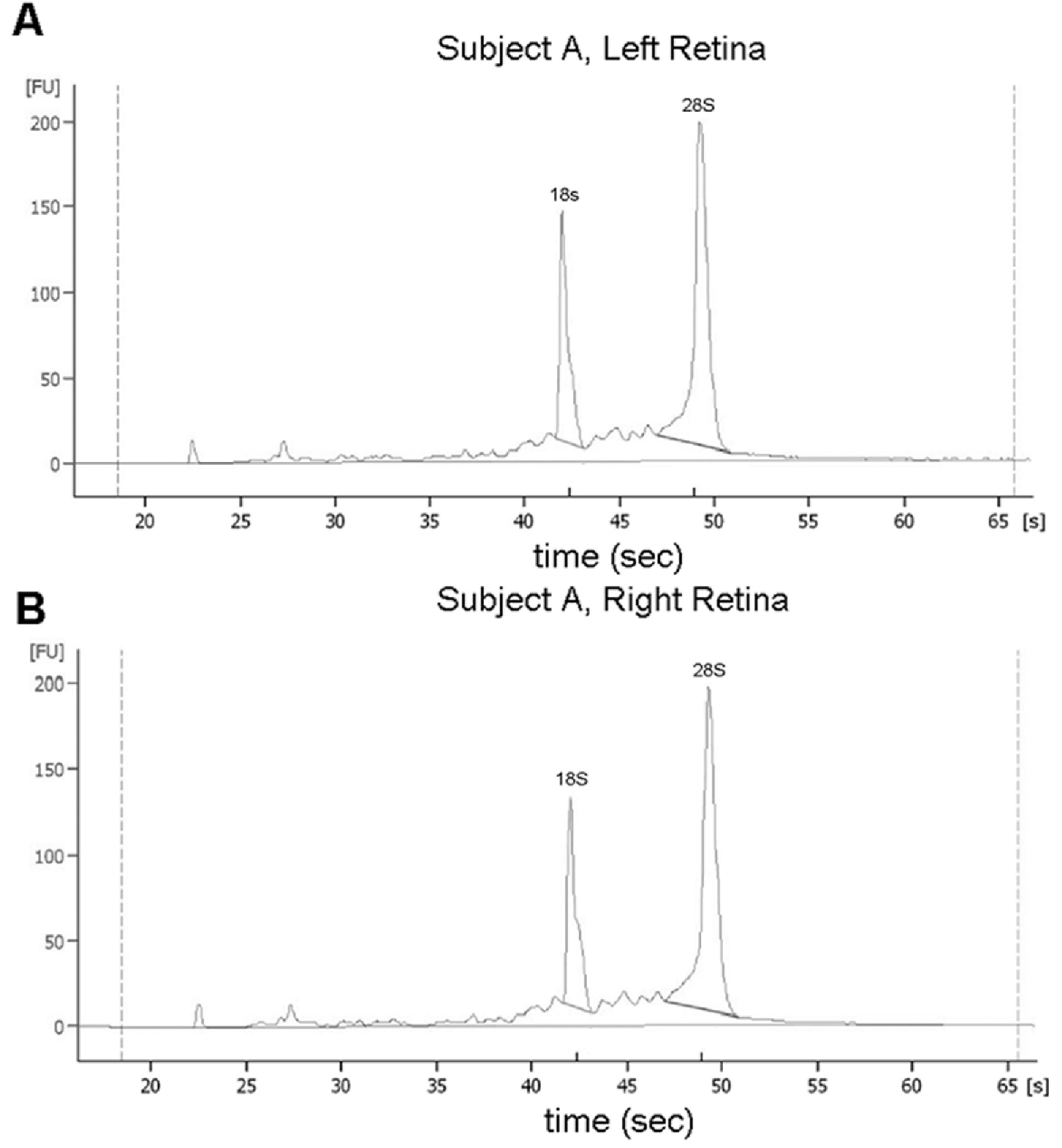
Figure 3. Electropherograms of total RNA
extracted from the retinas of left and right marmoset eyes of subject A
following microcapillary electrophoresis. Electropherograms demonstrate
well defined 18S and 28S rRNA peaks indicating high quality RNA was
extracted from the retinas of left (A) and right (B)
marmoset eyes. Since the retinas were dissected and processed in
parallel with the choroid/RPE tissue used in the present study, these
results suggest that the tissue dissection, snap freezing, and shipping
of tissues was adequate to preserve high quality RNA. FU is
fluorescence units.