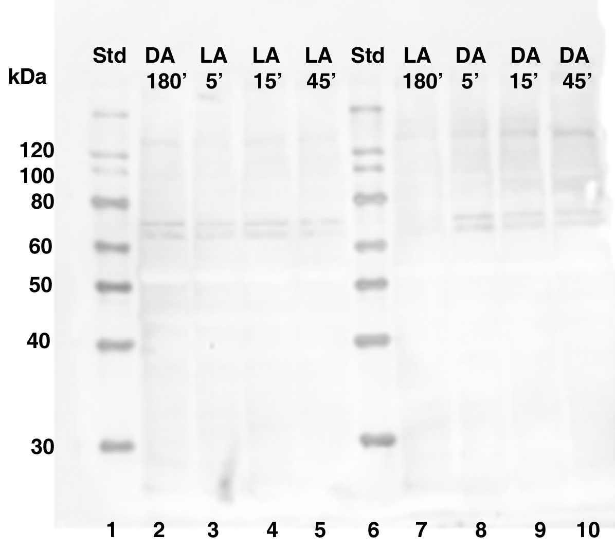
Figure 6. Western blot showing the effect
of inhibition of the CPEB antibody with PEP-171 neutralizing peptide.
Equal concentrations of total retinal protein was isolated from control
octopi at 180 min of dark or light adaptation and from octopi moved to
the opposite lighting conditions at 5 min, 15 min, and 45 min time
intervals. Lanes 2-5 and 7-10: The signal of the two major
immunoreactive bands present between 60-80 kDa is remarkably reduced,
as is the signal of smaller band is detected below 30 kDa. The
molecular weight sizes correspond to the fragments of the Magic Mark XP
protein standard (Invitrogen) in lanes 1 and 6. The following
abbreviations are used in the figure: standard (Std), light-adapted
(LA), and dark-adapted (DA).