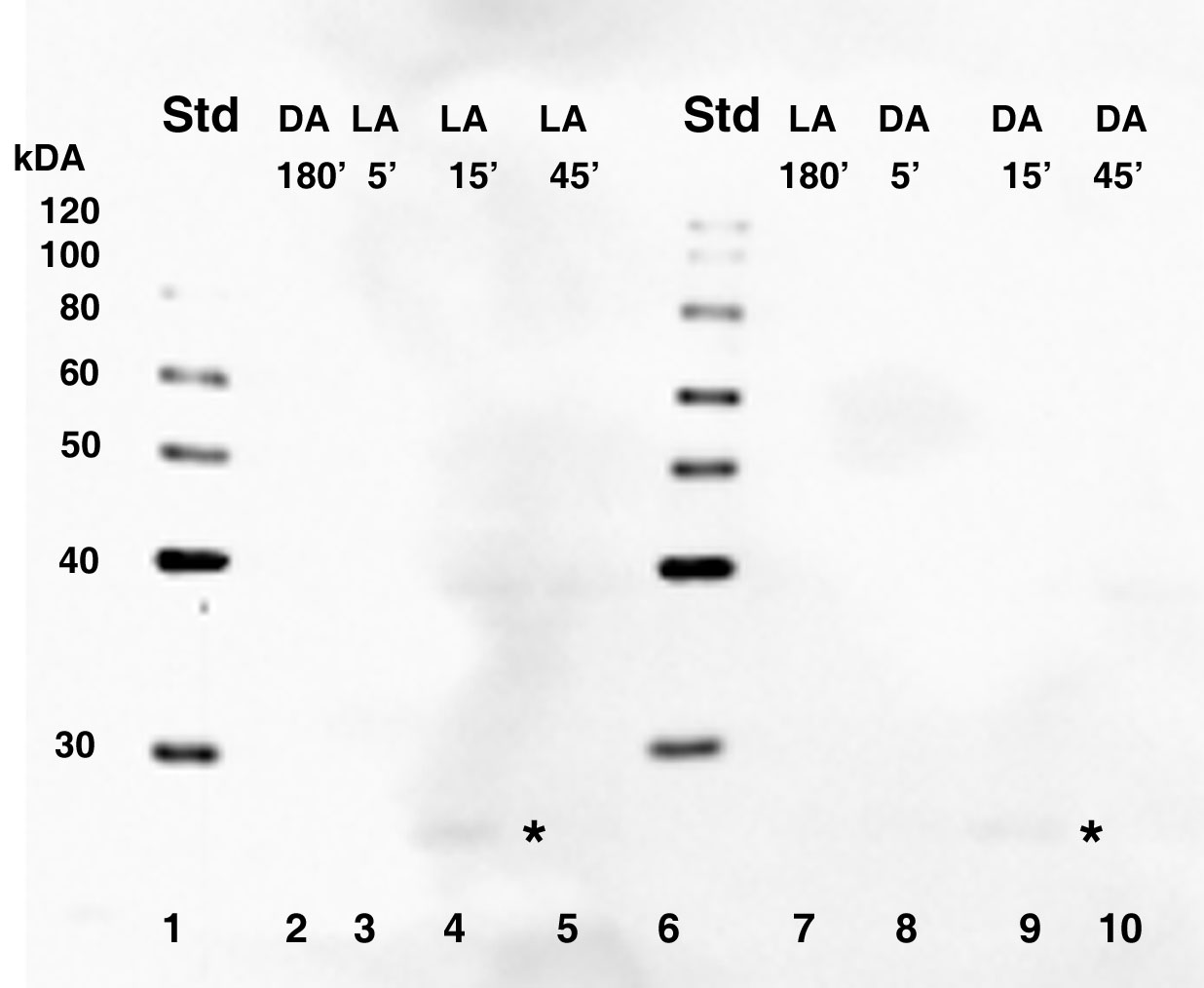
Figure 5. Western blot showing secondary
GAR-HRP antibody detection only. The detection of CPEB in dark-adapted
(DA) and light-adapted (LA) retinas does not result from nonspecific
antibody binding. Equal concentrations of total retinal protein was
isolated from control octopi at 180 min of dark or light adaptation and
from octopi moved to the opposite lighting conditions at 5 min, 15 min,
and 45 min time intervals. Lanes 2-5 and 7-10: The signals of the two
major immunoreactive bands present between 60-80 kDa were not detected.
Also slightly visible is a third smaller signal migrating below 30 kDa
(asterisk). The molecular weight sizes correspond to the fragments of
the Magic Mark XP protein standard (Invitrogen) in lanes 1 and 6. In
the figure, standard is abbreviated Std.