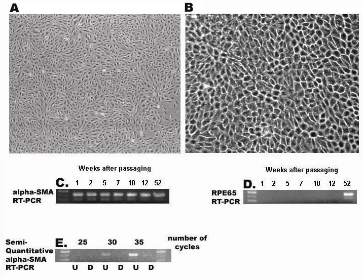
Figure 10. Reverse transcriptase
polymerase chain reaction analysis of markers during retinal pigment
epithelium cell differentiation. mRNA was isolated from
undifferentiated (A) or differentiated (B) ARPE-19 cells
and subjected to reverse transcriptase polymerase chain reaction
amplification to detect SMA (C) or RPE65 (D) as described
in Methods. Phase-contrast micrographs represent undifferentiated (A)
or differentiated (B) ARPE-19 cells after one week (A) or
52 weeks (B) of culture. C and D represent
RT–PCR amplification of mRNA samples isolated from ARPE-19 cells
maintained in culture for 1, 2, 5, 7, 10, 12, and 52 weeks, using
primers to detect mRNA for either αSMA (C) or RPE65 (D). E
represents RT–PCR amplification for a series of 25, 30, or 35 cycles to
detect αSMA using mRNA isolated from ARPE-19 cells that are
undifferentiated (U) differentiated (D). The first lane in C-E
represents a DNA standard ladder of 300, 400, and 500 bp.