Figure 2. Colocalization of TGF-β-RII and
PECAM-1. Native conjunctiva (A) and scarred filtering bleb
specimens (B, C) were double-stained for TGF-β-RII
(green) and the vascular endothelial cell marker, PECAM-1 (red). C
is a close-up of a section of B as indicated by the frame.
Arrows point to elongated TGF-β-RII positive cells devoid of PECAM-1
signal, which are most likely fibroblasts. Scale bar represents 50 µm (A,
B) and 10 µm (C).
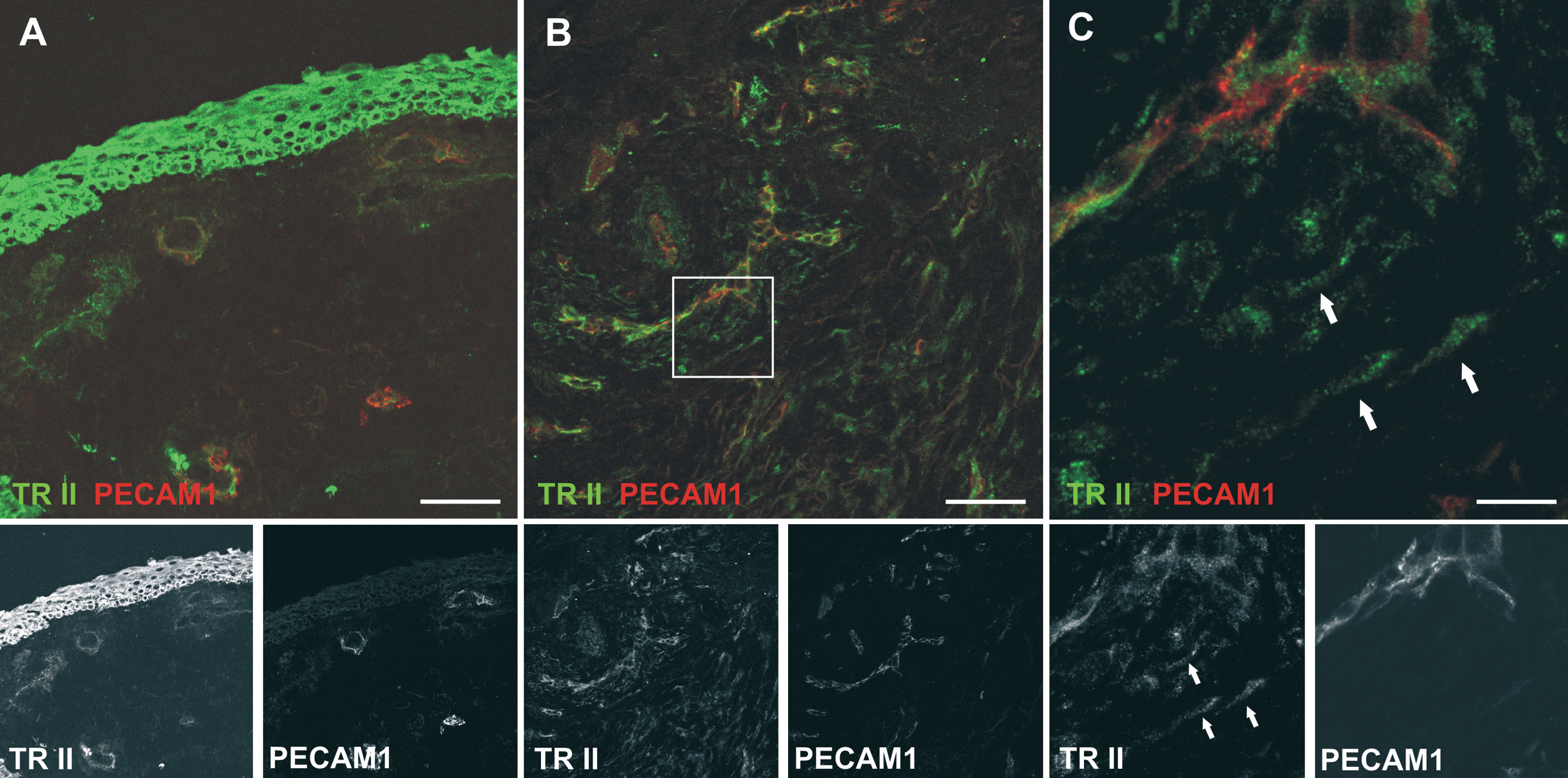
 Figure 2 of Meyer-ter-Vehn, Mol Vis 2008; 14:136-141.