Figure 1. Increased cell density in
scarred filtering blebs. Normal conjunctiva (A) shows loose
subepithelial connective tissue with sparse fibroblasts and occasional
vascular structures (arrows). In contrast, scarred filtering blebs (B)
are characterized by compacted connective tissue with abundant
spindle-shaped cells and various vascular structures. H&E stains.
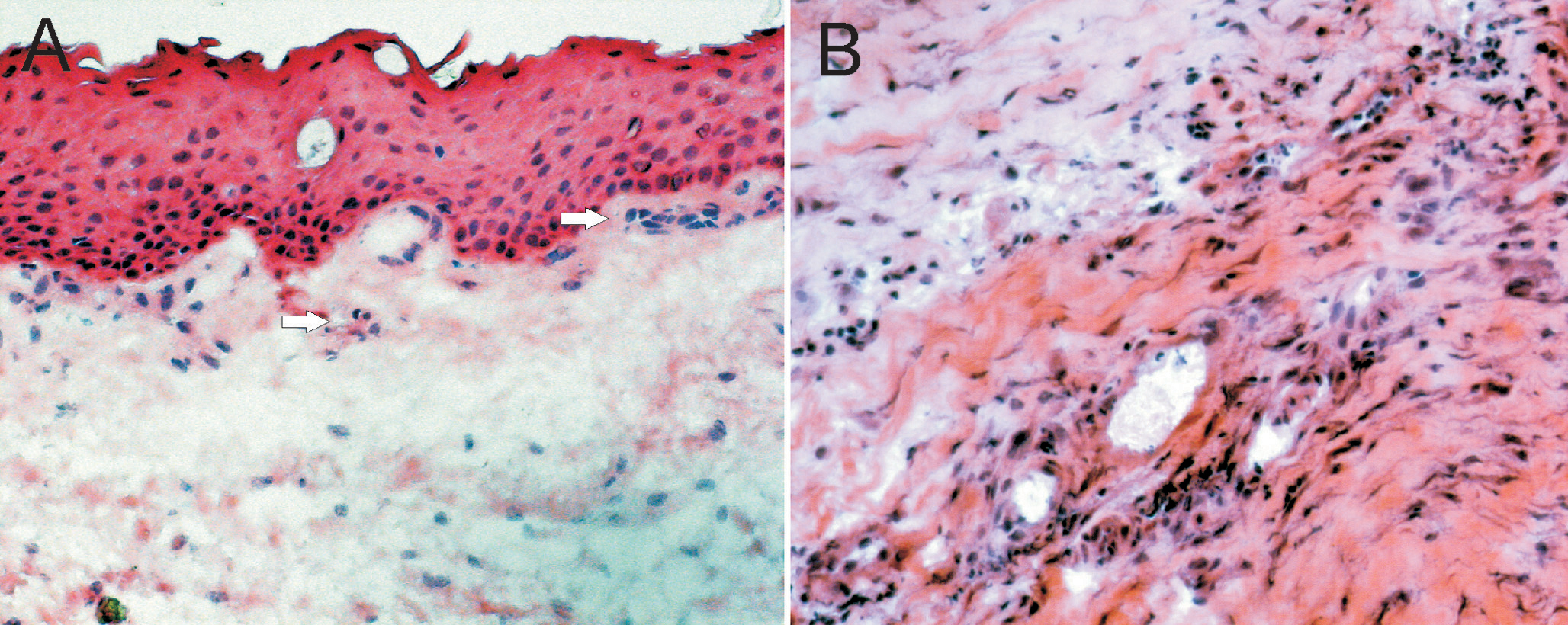
 Figure 1 of Meyer-ter-Vehn, Mol Vis 2008; 14:136-141.