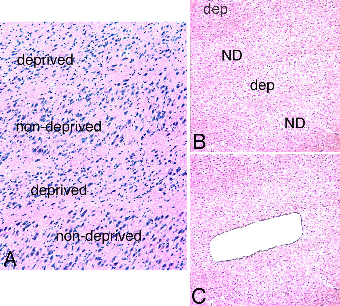
Figure 1. Morphology of LGN parvocellular
layers from a monkey with monocular vision deprivation. A:
H&E staining of lateral geniculate nucleus (LGN) sections showed
neuronal shrinkage in deprived parvocellular layers as compared with
non-deprived layers. B: Low power magnification deprived layer
of LGN sections before laser capture microdissection (LCM). C:
Low power magnification deprived layer of LGN sections after LCM.
Abbreviations: dep: deprived layer; ND: non-deprived layer.