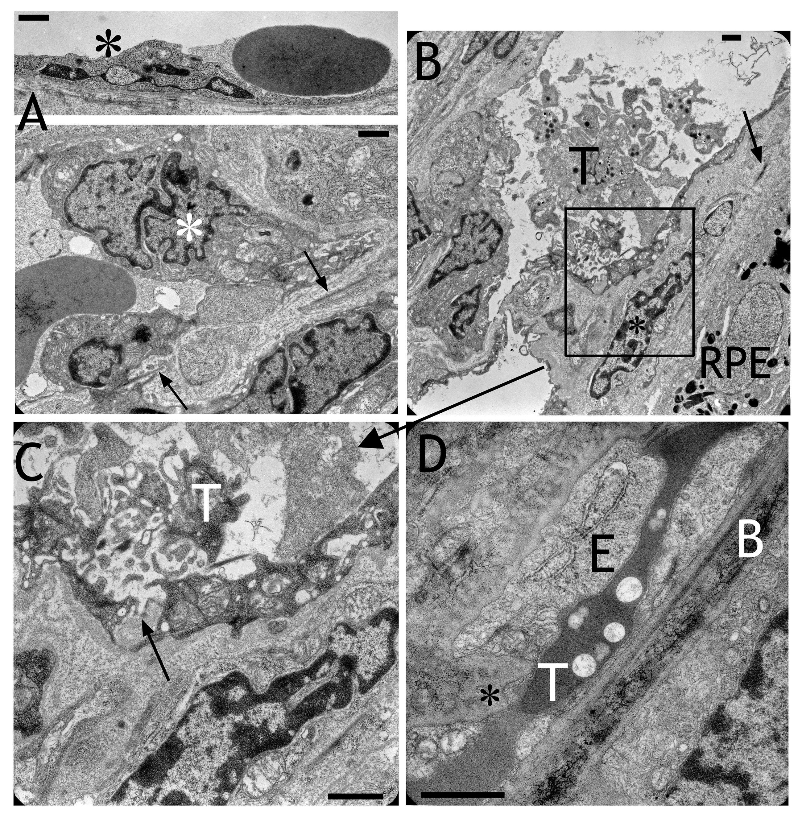
Figure 8. Electron micrographs obtained
after transduction with HC Ad.VEGF-A. A: Nuclei of the
choriocapillaris appeared extremely frayed or fragmented (asterisks).
Remnants of the elastic layer of Bruch’s membrane can be recognized
(arrows). B: Trombocytes (T) aggregated in the
choriocapillaris. Cells (black asterisk) migrated into Bruch’s membrane
between RPE and choriocapillaris. The elastic layer is marked by an
arrow. C: Magnification of the frame in (B) showed that
the adherent junctions were opened and the extracellular matrix had
direct contact with the lumen of the capillary (arrow). D: The
choriocapillaris was completely closed by thrombi (T), which was
directly faced Bruch’s membrane (B). The endothelium (E) was thickened
and interrupted by another cell (black asterisk). Scale bars in each
image equal 1 μm.