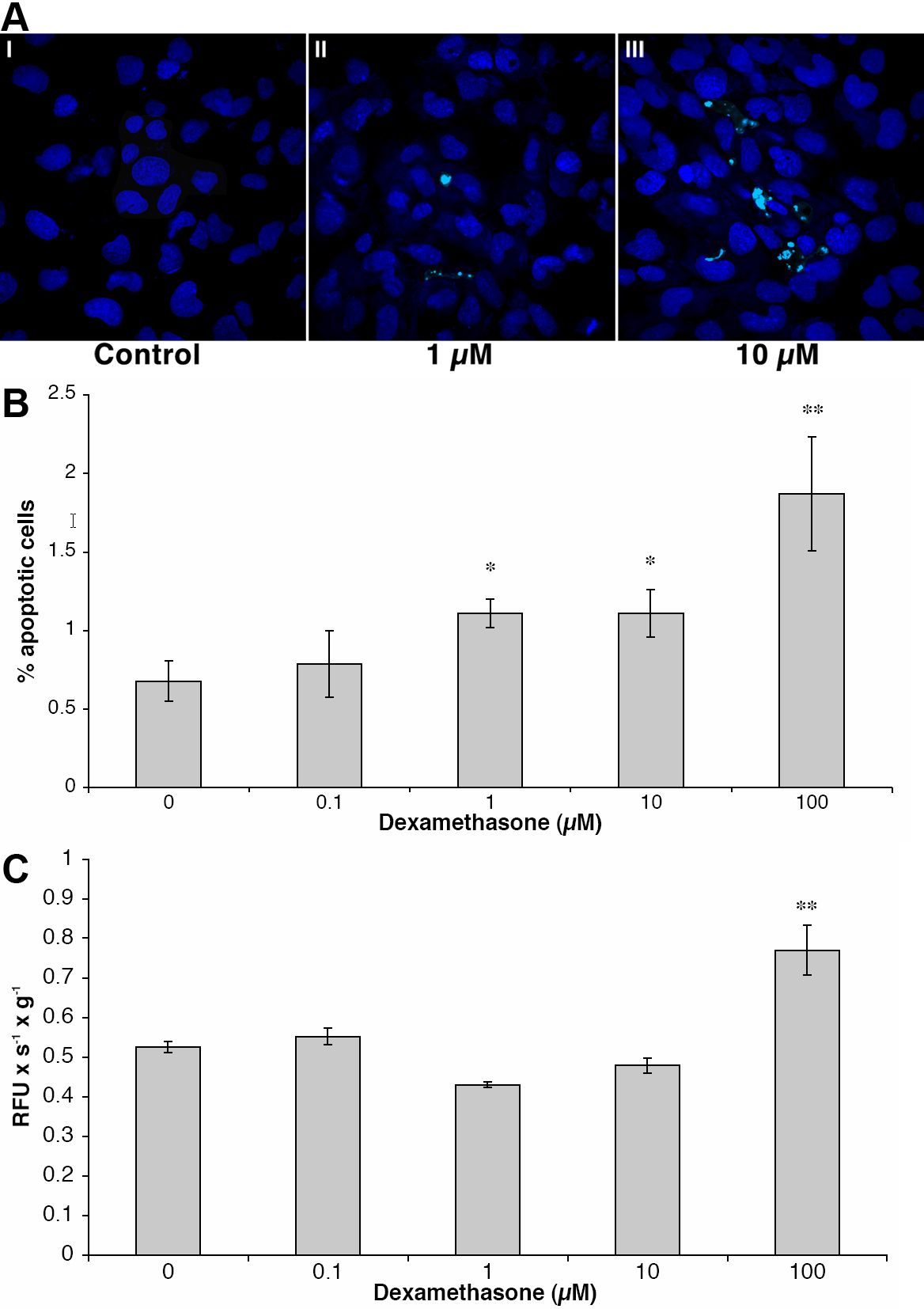
Figure 3. A: Apoptotic morphology
of human lens epithelial cells after dexamethasone exposure. Cells that
were exposed to dexamethasone exhibited morphologic changes typical of
apoptosis such as shrinkage, chromatin condensation, and nuclear
fragmentation (II and III). Cultured human lens epithelial cells are
shown stained with the nuclear dye Hoechst 33342. Control cells (I) and
cells exposed to 1 µM (II) and 100 µM (III) dexamethasone are shown.
Original magnification 600X. B: Showing increased number of
apoptotic cells after dexamethasone exposure. Human lens epithelial
cells were exposed to dexamethasone at different concentrations during
24 hs after which the cell nuclei were stained with Hoechst 33342. The
percentage of apoptotic cells increased in a dose-dependent manner with
higher dexamethasone concentrations. At least 300 cells from three
different chamber slides were counted. Mean ±SEM are given; the
asterisk indicates a p<0.05 and the double asterisk indicates a
p<0.01. C: Increased Caspase-3 activity after incubation
with dexamethasone. The caspase-3 activity in cultured human lens
epithelial cells was significantly increased after the administration
of 100 µM dexamethasone for 24 h. A representative experimental
run from three experiments with similar results is shown. Caspase-3
activity is expressed as relative fluorescence units per second and
gram protein (RFU s−1g−1). Mean ±SEM from 3
separate culture wells are shown; the double asterisk indicates a
p<0.01.