![]() Figure 2 of
Schmidt, Mol Vis 2008;
14:125-135.
Figure 2 of
Schmidt, Mol Vis 2008;
14:125-135.
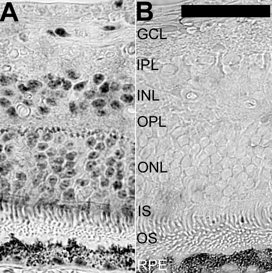
Figure 2. Immunostaining for 110 kDa SR-related protein in human retina
Immunohistochemical detection of the 110 kDa SR-related protein of the U4/U6.U5 tri-snRNP (hypoxia-associated factor) was carried out in paraffin sections of the peripheral human retina. The dark signal in the retinal pigment epithelium (RPE) and choroid derives from the melanin. These areas were included in the image to illustrate the total absence of specific immunolabeling from the outer segments. A: Distinct cellular labeling was obtained with the antibody to 110 kDa SR-related protein of the U4/U6.U5 tri-snRNP. B: Immunoreactivity was absent after absorption of the primary antibody with immunogenic peptides. The following abbreviations were used in this figure: ganglion cell layer (GCL), inner nuclear layer (INL), inner plexiform layer (IPL), outer plexiform layer (OPL), inner segments of photoreceptors (IS), outer nuclear layer (ONL), and outer segments of photoreceptors (OS). The calibration bar is equal to 40 μm.