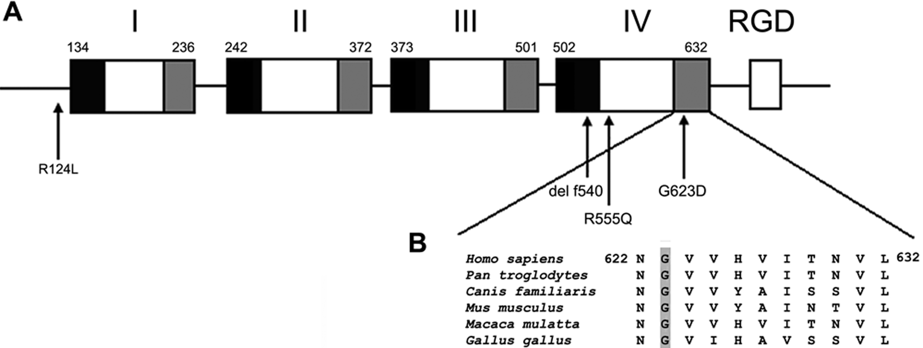
Figure 4. Schematic diagram of TGFBI. A:
A diagram of recombinant ßIG-H3 proteins is illustrated. Black and gray
boxes indicate the highly conserved sequences of each repeated domain.
The RGD motif is shown as an open box. Mutations of TGFBI
associated with RBCD occur at amino acids 124, 555, 540, and 623 are
indicated by arrows. B: The alignment of TGFBI
sequences in diverse species is shown. The glycine is conserved in
TGFBI proteins from several species.