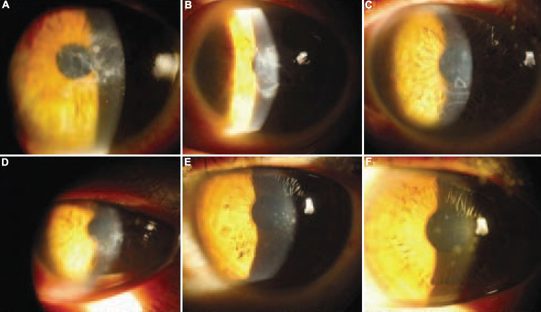
Figure 2. Clinical photography of corneas
in affected family members. A and B: Slit lamp
photographs of the proband show multiple opacities in the subepithelial
and anterior stromal regions in the central cornea of the left eye (A)
and geographical opacity involving the anterior and mid stroma in the
right eye (B). C and D: Corneal images of
patient IV:3 revealed geometric and round opacities involving the
anterior stroma and subepithelial layers in both eyes. E and F:
Corneal images of V:1 and V:3 are shown, respectively. Dot epithelial
and subepithelial opacities were noted.