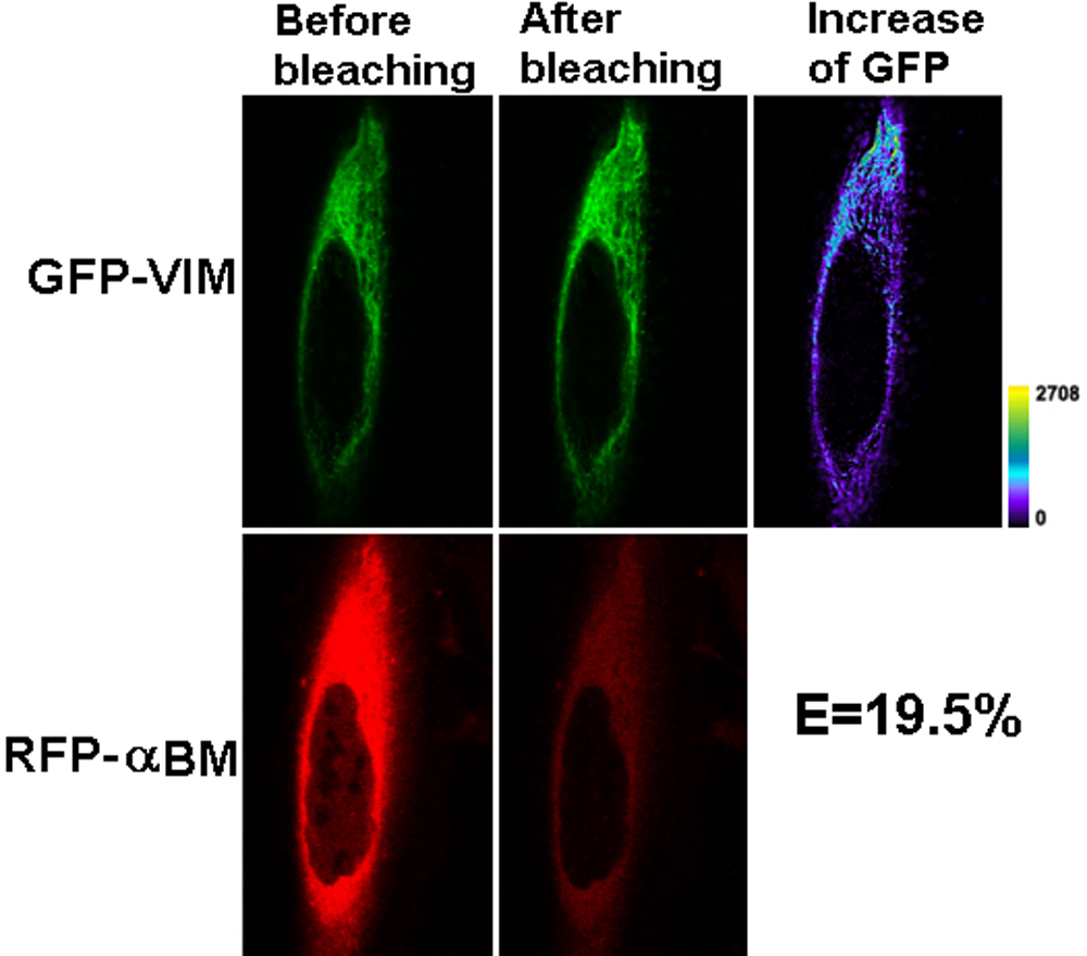
Figure 5. Representative laser scanning
microscopy images of FRET acceptor photobleaching of HeLa cells
co-transfected with GFP-VIM and RFP-R120G αB. The pair of constructs
was co-transfected into HeLa cells. After culture, laser scanning
microscopy (LSM) images were taken before and after photobleaching of
the acceptor for 45 s with a 543 nm laser beam. A decrease of red
fluorescence and increase of green fluorescence were observed. The
transfer efficiency was calculated with the equation: E=1 – FGFP-min/FGFP-max.
The efficiency for this cell that co-transfected with GFP-VIM and
RFP-R120G αB is 19.5%, twofold greater than the GFP-VIM and RFP-WTαB.
The increase of GFP fluorescence intensity is converted to pseudocolor
(right panel) that displays variations of pixel gray scales with color.