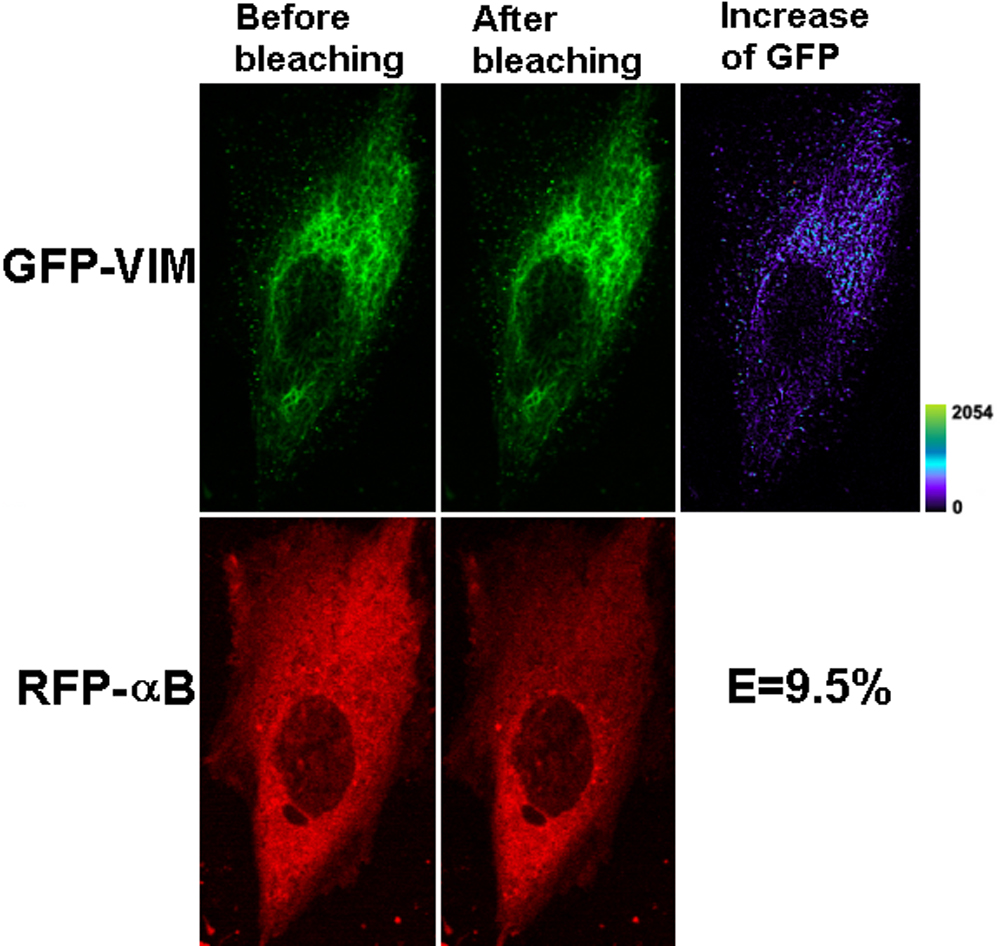
Figure 4. Representative laser scanning
microscopy images of FRET acceptor photobleaching of HeLa cells
co-transfected with GFP-VIM and RFP-WTαB. The pair of constructs was
co-transfected into HeLa cells. After culture, laser scanning
microscopy (LSM) images were taken before and after photobleaching of
the acceptor for 45 s with a 543 nm laser beam. A decrease of red
fluorescence and increase of green fluorescence were observed. The
transfer efficiency was calculated with the equation: E=1 – FGFP-min/FGFP-max..
The efficiency for this cell that co-transfected with GFP-VIM and
RFP-WTαB is 9.5%, much greater than the negative control of untagged
GFP and RFP. The increase of GFP fluorescence intensity is converted to
pseudocolor (right panel) that displays variations of pixel gray scales
with color.