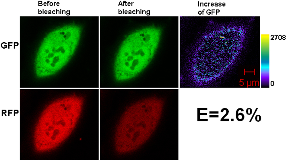
Figure 3. Representative laser scanning
microscopy images of HeLa cells co-transfected with the negative
controls, untagged GFP and RFP. The pair of constructs was
co-transfected into HeLa cells. After culture, laser scanning
microscopy (LSM) images were taken. The low efficiency shown arises
from experimental background. The increase of GFP fluorescence
intensity is converted to pseudocolor (right panel) that displays
variations of pixel gray scales with color.