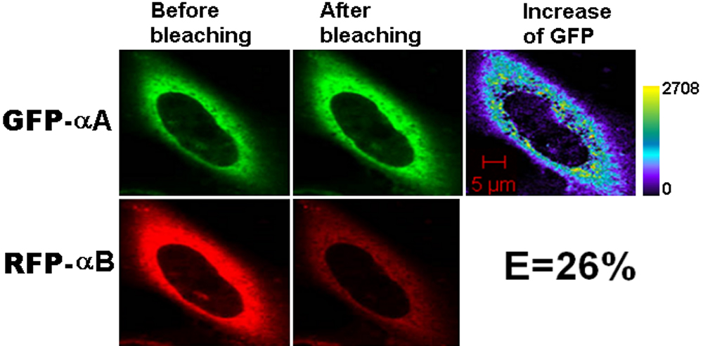
Figure 2. Representative laser scanning
microscopy images of HeLa cells co-transfected with the positive
controls, GFP-αA and RFP-αB. The constructs were co-transfected into
HeLa cells. After culture, laser scanning microscopy (LSM) images were
taken. αA- and αB-crystallins are known to have a strong
subunit-subunit interaction. The energy transfer efficiency is high.
The increase of GFP fluorescence intensity is converted to pseudocolor
(right panel) that displays variations of pixel gray scales with color.

![]() Figure 2 of Song, Mol Vis 2008; 14:1282-1287.
Figure 2 of Song, Mol Vis 2008; 14:1282-1287. 