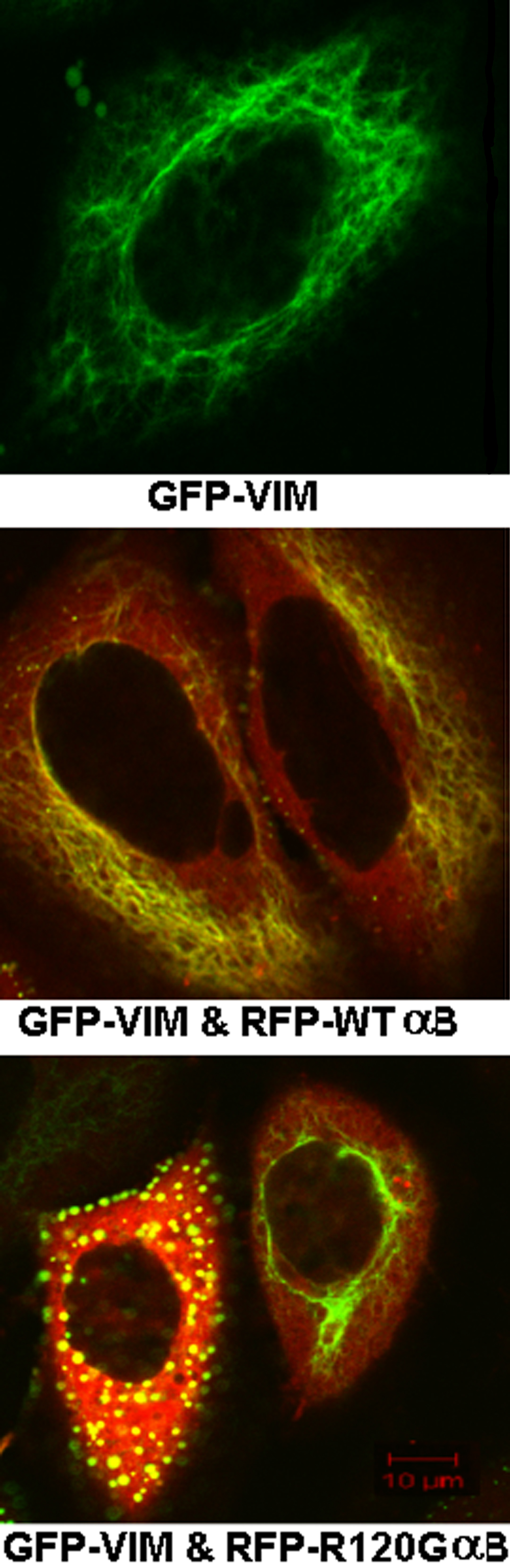
Figure 1. Representative laser scanning
microscopy images of HeLa cells transfected with GFP-VIM and
co-transfected with GFP-VIM and WT αB-crystallin (αB) or R120G
αB-crystallin (αBM). The single construct or pair of constructs was
transfected into HeLa cells. After culture, laser scanning microscopy
(LSM) images were taken. Either the green image (GFP-VIM) or merged
image of green and red fluorescence (GFP-VIM and RFP-WT αB-crystallin
[αB] or GFP-VIM and RFP-R120G αB) was shown. Vimentin filaments are
clearly shown in cells co-expressing WT αB-crystallin, but enormous
aggregates were formed in the cells co-expressing R120G αB-crystallin.
Vimentin filaments are shown as the fibrous structures and aggregates
as the bright, dense spots.