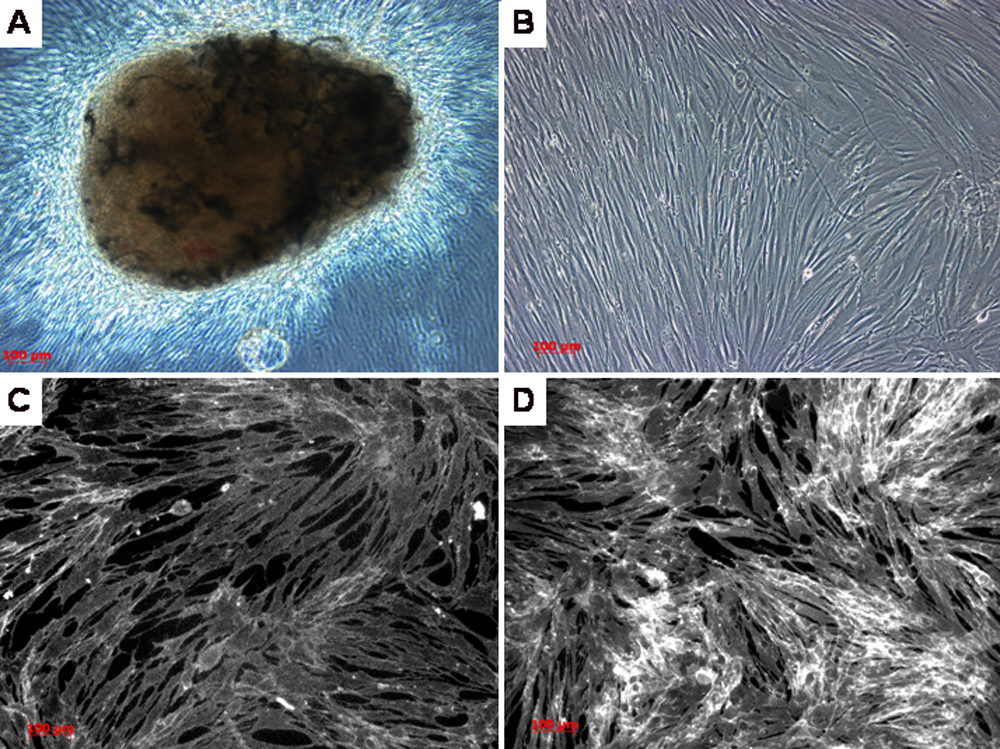
Figure 5. Fibroblast cell culture from the
conjunctival biopsy of the proband. A: Fibroblastic cellular
outgrowth from the conjunctival biopsy tissue. B: Following one
week of cell culture, cellular confluence of fibroblasts is achieved. C:
Following the addition of filipin stain, there is no evidence of
cholesterol in the cultured conjunctival fibroblasts. D: Cells
cultured with the addition of low density lipoprotein (LDL), acted as a
positive control showing evidence of cholesterol with filipin staining.
(Bar=100 microns).