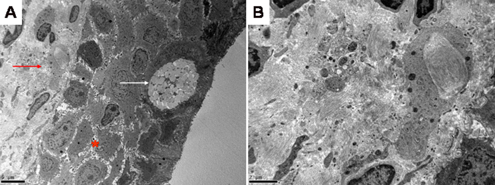
Figure 4. Transmission electron microscopy
of conjunctival biopsy from the proband. A: Low power (2000X)
section showing conjunctival epithelium (red star), goblet cell (white
arrow) and conjunctival fibroblast (red arrow). B: High
power section (5000X) of stromal fibroblastic tissue showing an absence
of lipids in the cytoplasm of the conjunctival fibroblast.