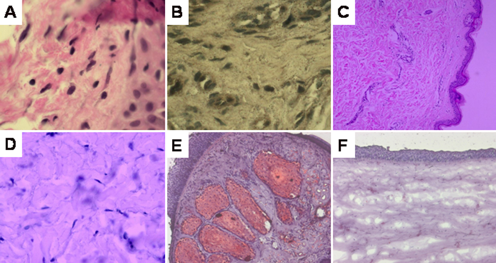
Figure 3. Histological sections of
conjunctival and skin biopsy, from the proband. A: Hematoxylin
and Eosin staining of conjunctival fibroblasts (40X). B:
Negative result of oil Red-O staining of conjunctival fibroblasts
(400X). C: Hematoxylin and Eosin staining of skin biopsy (40X).
D: Negative result of oil Red-O staining
of dermal fibroblasts (400X). E: Positive control for
oil Red-O stain; lid margin staining the meiobium gland (100X). F:
Negative control for oil Red-O stain; Normal cornea (100X).