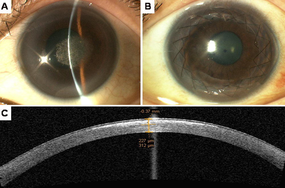
Figure 1. Color slit lamp photomicrographs
of the proband IV:4. A: The image shows the proband’s left eye
(OS) before ALTK. B: The image shows the same eye five years
following ALTK with no signs of recurrence. C: Pre
Pre-operative ASOCT of the proband showing hyper-reflective signal,
indicating the depth of the crystalline lesions.