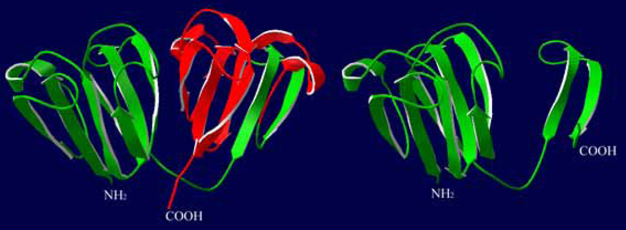
Figure 6. Comparative modeling of the full
length and truncated γC-crystallins. The structural modeling was
analyzed in Swiss-PdbViewer (version 3.7). When comparing the full
length (left) and truncated γC-crystallins (right), the six
COOH-terminal β-strands are truncated in the mutated γC-crystallin (the
strands are shown in red).